

Amyloid Beta Aggregation Protocol
|
This detailed protocol provides everything you need to successfully work with and aggregate Amyloid Beta (Aβ) for your experiments in-vitro and in-vivo. Written by our PhD qualified expert team, this protocol includes help with handling Aβ monomers, tips on monitoring Aβ aggregation and protocols for generating Aβ fibrils of different sizes. |
 |
1. Background
Amyloid Beta (Aβ) is a family of peptides that form the main constituent of senile plaques in Alzheimer's disease and has been extensively investigated for its role in the aetiology of neurodegenerative diseases. Aβ is produced from the proteolytic cleavage of amyloid precursor protein (APP), a membrane glycoprotein found extensively in synapses throughout the brain (see Figure 1) to result in a family of peptides ranging in size from 37 to 49 amino acids long with the main species being Aβ1-40 and Aβ1-42.
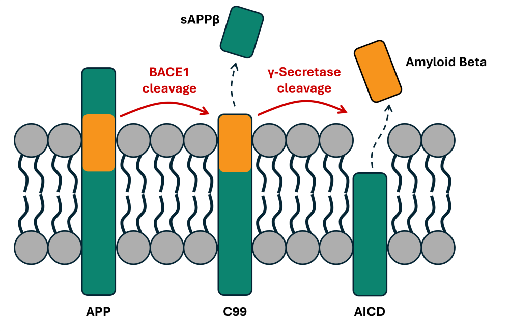
Figure 1. Generation of amyloid beta through APP proteolytic cleavage. Amyloid precursor protein (APP) is cleaved by β-secretases such as BACE1 to give a 99 residue C-terminal fragment (C99) which is then cleaved again by the γ-secretase complex to liberate free amyloid beta which mostly takes the form of either Aβ1-40 or Aβ1-42.
Monomeric Aβ has multiple roles in normal physiology where it is involved in trafficking of synaptic vesicles, regulating the excitation / inhibition balance at synapses and activating pathways regulating BDNF release. However Aβ isoforms are prone to aggregation where they form larger and larger complexes and aggregates which are the main driver of their toxicity. Aβ monomers start to coalesce into small soluble oligomers (complexes of 2-20 monomers) which are now believed to be the major toxic species and can diffuse throughout the brain. Aβ oligomers have an array of toxic effects including disruption of synaptic plasticity, promotion of tau hyperphosphorylation and triggering of oxidative stress. Oligomers can coalesce into soluble protofibrils which have additional toxic effects such as being endocytosed by glial cells and being associated with inflammation. These protofibrils can then further aggregate into insoluble fibrils which have a characteristic cross-β sheet pattern which then further accumulate into the plaque deposits found in the brains of patients with Alzheimer's disease.
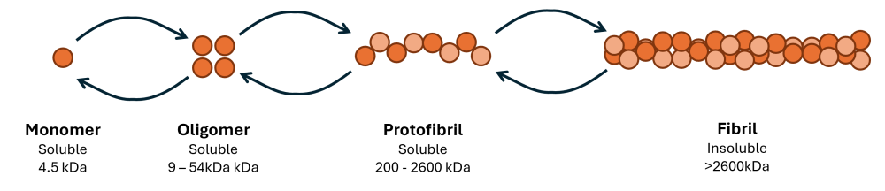
Figure 2. Aggregation of soluble Aβ monomers into insoluble fibrils is a dynamic and concentration dependent process in equilibrium.
There are a range of different Aβ species but the most widely studied in neurodegenerative research are:
- Amyloid β 1-40 - comprises about 90% of the total Aβ formed in the brain and while more soluble than Aβ1-42 still forms a major constituent of senile plaques in Alzheimer's disease.
- Available from Hello Bio as HB8758 - β-Amyloid Peptide (1-40) (human), a high purity lyophilized monomer in a 1mg pack size.
- Amyloid β 1-42 - comprises only around 10% of Aβ in the brain, however it is less soluble, more neurotoxic and quicker to aggregate than Aβ1-40 due to a higher propensity to form β-sheet structures.
- Available from Hello Bio as HB9805 - β-Amyloid Peptide (1-42) (human), a high purity lyophilized monomer in packs ranging from 100µg to 1mg.
- Amyloid β 25-35 - produced from the cleavage of Aβ1-40 and is another component of senile amyloid plaques in the brain. Aβ25-35 is also able to aggregate into fibrils and has been found to have cytotoxic effects.
- Available from Hello Bio as HB8524 - β-Amyloid Peptide (25-35) (human), a high purity lyophilized monomer in a 1mg pack size.
- Amyloid β 42-1 - a reverse form of the Aβ1-42 peptide that is used as a control peptide in experiments using Aβ1-42 as, while it has been found to aggregate, it is much less toxic to cells than Aβ1-42
- Available from Hello Bio as HB9159 - β-Amyloid Peptide (42-1) (human), a high purity lyophilized monomer in a 1mg pack size.
2. Handling Monomers
Amyloid β monomers supplied by Hello Bio are provided as a lyophilizate that has been previously treated with HFIP. We recommend to dissolve peptides following the below NH4OH protocol although others are provided in section 3.1.
- Dissolve Aβ peptide in 1% ammonium hydroxide (NH4OH) using a volume of around 70-80µl per 1mg of peptide. Briefly spin the vial at >1000g to bring all the solution to the bottom of the vial.
- 1% ammonium hydroxide is conveniently available preformulated from Hello Bio as HB9790 - 1% NH4OH solution for dissolving Beta-Amyloid peptides
- Do not store the peptide in 1% NH4OH
- Immediately dilute the Aβ solution in ice-cold PBS to a concentration of ≤1mg/ml. Pipette up and down to ensure the solution is well mixed.
- Either use immediately or snap-freeze the solution in aliquots and store at -80°C.
- Before use it is recommended to spin the Aβ aliquot at full speed in a benchtop centrifuge to remove any small insoluble aggregates that have formed.
We recommend following these tips to avoid undesired aggregation of Aβ peptides:
- Use clean tools to avoid introducing contaminants into the solution that could be seeds for aggregation.
- Avoid extensively vortexing Aβ samples as this can promote aggregation.
- Be careful to avoid introducing bubbles into Aβ solutions as this can be another source of seeds for aggregation.
- Avoid freeze-thaw of Aβ solutions as this has been found to promote aggregation.
3. Aggregation of Amyloid Beta
There are a diverse range of methods used to generate different oligomerisation species of Aβ each with their own range of advantages and disadvantages. We have a protocol based upon the work of Stine et al., 2011. It is important to start with a solution of pure Aβ monomers to maximize the probability of resulting in a uniform aggregation state at the end.
3.1 Starting with Pure Monomeric Aβ
There are a few different methods that are used to ensure a solution of pure monomeric Aβ. Please note: if planning on oligomerizing Aβ then we recommend dissolving straight from the vial with DMSO (please see section 3.3).
HFIP Treatment
- Dissolve the Aβ peptide in HFIP (1,1,1,3,3,3-hexafluoro-2-propanol, CAS: 920-66-1) and vortex briefly to mix.
- Dry the solution under a stream of nitrogen
- Redissolve the peptide in HFIP at 1mg/ml, sonicate in a bath sonicator for 5 minutes.
- Dry the solution under a stream of nitrogen
- Redissolve the peptide in HFIP at 1mg/ml, sonicate in a bath sonicator for 5 minutes.
- Dry the solution under a stream of nitrogen
- Redissolve the peptide in HFIP at 1mg/ml, aliquot into smaller volumes appropriate for a single experiment then sonicate in a bath sonicator for 5 minutes.
- Dry the solution under a stream of nitrogen followed by drying under vacuum for 1-2 hours until the peptide is completely dry.
- Store the Aβ aliquots at -80°C until further use.
NaOH Treatment
- Dissolve the Aβ peptide in 10mM NaOH to 1mg/ml and sonicate for 30 minutes using a sonicator bath.
- Aliquot and snap freeze before storing at -80°C
- When ready to use, thaw rapidly at 37°C then remember to account for the high pH of the Aβ solution when using it.
NH4OH Treatment
- Dissolve Aβ peptide in 1% ammonium hydroxide (NH4OH) using a volume of around 70-80µl per 1mg of peptide. Briefly spin the vial at >1000g to bring all the solution to the bottom of the vial.
- 1% ammonium hydroxide is conveniently available preformulated from Hello Bio as HB9790 - 1% NH4OH solution for dissolving Beta-Amyloid peptides
- Do not store the peptide in 1% NH4OH
- Immediately dilute the Aβ solution in ice-cold PBS to a concentration of ≤1mg/ml. Pipette up and down to ensure the solution is well mixed.
- Either use immediately or snap-freeze the solution in aliquots and store at -80°C.
- Before use it is recommended to spin the Aβ aliquot at full speed in a benchtop centrifuge to remove any small insoluble aggregates that have formed.
3.2 How to Monitor Aggregation
Being able to monitor the progression of Amyloid Beta aggregation is critical to understand what species are being used in experiments where Aβ is aggregated then used as an in-vitro or in-vivo manipulation. There are multiple ways of measuring Aβ aggregation with it being recommended to use more than one method for a more accurate determination of Aβ aggregation status.
Fluorescent Assays
There are a range of fluorescent molecules that differentially bind to Aβ fibrils and fluoresce compared to monomers which are available from Hello Bio:
- HB5134 - Thioflavin S (ThS)
- HB17774 - Thioflavin X (ThX)
- HB7143 - Thioflavin T (ThT)
- HB5252 - Methoxy-X04
- HB0737 - Congo Red
- Prepare Aβ in 20mM phosphate buffer containing 0.2mM EDTA, 1mM NaN3 and 20µM Thioflavin T or 10µM Thioflavin X at pH 8. Aβ concentrations of over 1µM exhibit aggregation within a 24 hour timespan.
- Incubate for 24 hours at 37°C regularly measuring fluorescence at either:
- Thioflavin X: Excitation: 420, Emission: 494nm
- Thioflavin T: Excitation: 450nm, Emission: 485nm
Transmission Electron Microscopy (TEM)
Aβ fibrils are large enough to be visualized using transmission electron microscopy (TEM) with uranyl acetate counterstaining. A standard protocol from Xiao et al., 2015 is:
- Load 10µl of a fibril sample onto a 300 mesh copper formvar / carbon grid and incubate for 1 minute before blotting off any excess.
- Counterstain the sample with 10µl of 2% uranyl acetate for 90 seconds before blotting off any excess and drying the grid in a desiccator chamber.
- Image using a TEM at 80kV and a magnification of approximately 120,000x
Dynamic Light Scattering
Dynamic Light Scattering (DLS) is a technique that uses the scattering of a laser beam off small particles in solution to work out their size distribution (for an example see Jeon et al., 2023). This can be applied to monitoring Aβ aggregation as DLS is able to non-invasively measure Aβ aggregation in a timecourse and distinguish between small soluble oligomers and larger insoluble fibrils.
Atomic Force Microscopy (AFM)
Atomic Force Microscopy (AFM) is a technique where a nanoscale probe is dragged across a sample then the deflection measured to map the surface topology of a sample. This has been successfully applied to monitor the aggregation of Aβ protofibrils into fibrils (see Harper et al., 1997) and can directly visualize the structure of aggregated Aβ and even has the resolution to visualize the helical structure of mature fibrils.
3.3 Aggregation Protocols
Depending on the size of aggregate you require for your experiments there are different protocols which bias their conditions towards making fibrils assemble to a certain range of sizes. We recommend in all cases dissolving in DMSO straight from the vial for aggregating Aβ.
3.3.1 Preparing Oligomers
- Dissolve monomeric Aβ in DMSO to a 5mM concentration at room temperature (22.6mg/ml for Aβ1-42)
- Dilute the Aβ down to 100µM (0.45mg/ml for Aβ1-42) using ice-cold F-12 cell culture media (phenol free and containing 146mg/L L-glutamine)
- Vortex for 15 seconds then incubate at 4°C for 24 hours.
3.3.2 Preparing Fibrils
- Dissolve monomeric Aβ in DMSO to a 5mM concentration at room temperature (22.6mg/ml for Aβ1-42)
- Dilute the Aβ down to 100µM (0.45mg/ml for Aβ1-42) using room temperature 10mM HCl.
- Vortex for 15 seconds then incubate at 37°C for 24 hours.
3.3.3 Preparing Larger Insoluble Aggregates
- Dissolve monomeric Aβ in DMSO to a 5mM concentration at room temperature (22.6mg/ml for Aβ1-42)
- Dilute the Aβ down to 100µM (0.45mg/ml for Aβ1-42) using room temperature 10mM HCl + 150mM NaCl.
- Vortex for 15 seconds then incubate at 37°C for 24 hours.
3.4 Separating Aβ Aggregates
Depending on the experimental requirements, the Aβ aggregate mixture can be used directly or purified to isolate specific species. There are a few different methods used to separate Aβ species following an aggregation protocol:
- Filtration is an easy method to remove larger Aβ fibrils and aggregates with spin-column filters being available with a wide range of molecular weight cut offs (MWCO), ranging from as little as 1kDa up to over 100kDa. Spin filtration is extremely quick, however is relatively crude as it only removes Aβ species above the MWCO, leaving a mixture of smaller species remaining.
- Size exclusion chromatography (SEC, see Nichols et al., 2015) is the main method used to separate Aβ species where Aβ species are separated by hydrodynamic size as they move through the resin bed with larger species eluting before smaller oligomers and monomers. SEC is an extremely powerful technique however does require expensive columns and machinery to carry out.
- Aβ species can also be separated using centrifugation (see Ward et al., 2000). Mixed Aβ samples can be added to a density gradient and spun at extremely high g-force (approx. 350,000 gav) to separate the Aβ species by their molecular weight. This does however require expensive specialised centrifuges and long run-times making this method non-trivial.
4. Uses of Amyloid Beta Aggregates
Creation of high-purity and defined aggregation state amyloid beta species is critical to enable research into the specific molecular mechanisms underlying Aβ toxicity in-vivo which leads to Alzheimer's disease. Some of the most common uses for Aβ oligomeric species include:
- Screening for either small molecule and peptide inhibitors of Aβ aggregation or disaggregators of Aβ fibrils using high-throughput screening assays. Dyes such as Thioflavin X (ThX) and Thioflavin T (ThT) increase fluorescence in response to the formation of aggregated Aβ species therefore can be readily adapted into a high-throughput format.
- In-vitro toxicity assays where Aβ species are added to cells (e.g. primary or iPSC derived) neurons and the toxicological and molecular mechanisms of cell damage are assayed using tests such as H2DCFDA to measure reactive oxygen species, Annexin V assays to measure apoptosis or MTT to measure cell proliferation.
- HB7375 - DCFDA / H2DCFDA - Cellular ROS Assay Kit
- HB9623 - Annexin V-FITC Apoptosis Staining / Detection Kit
- HB8164 - Annexin V-PE Apoptosis Staining / Detection Kit
- HB5283 - MTT
- In-vivo models where animals are injected with toxic Aβ species to study their impact on a systems level. This is often carried out on genetically modified animals that overexpress human Aβ isoforms which then have Aβ fibrils injected to trigger further aggregation of host Aβ which produces an Alzheimer’s like phenotype in a greatly shortened timespan compared to the natural disease process. Researchers commonly carry out immunohistochemical analyses to identify the specific pathology on different cell types such as:
- Neurons (e.g. Anti-βIII Tubulin antibody ValidAb™, Anti-NeuN antibody ValidAb™, Anti-MAP2 antibody ValidAb™)
- Glia (e.g. Anti-GFAP antibody ValidAb™)
- Oligodendrocytes (e.g. Anti-Myelin Basic Protein (MBP) Antibody ValidAb™)
Fluorescent dyes can also be used to visualize Aβ plaques in histological samples from animal models such as:
- HB17774 - Thioflavin X (ThX) – next generation dye that is 5x brighter than ThT
- HB7143 - Thioflavin T (ThT)
- HB5134 - Thioflavin S (ThS)
- HB5252 - Methoxy-X04
- HB0737 - Congo Red
5. References
- Benilova I, Karran E, De Strooper B. The toxic Aβ oligomer and Alzheimer's disease: an emperor in need of clothes. Nat Neurosci. 2012 Jan 29;15(3):349-57. doi: 10.1038/nn.3028. PMID: 22286176.
- Chen GF, Xu TH, Yan Y, Zhou YR, Jiang Y, Melcher K, Xu HE. Amyloid beta: structure, biology and structure-based therapeutic development. Acta Pharmacol Sin. 2017 Sep;38(9):1205-1235. doi: 10.1038/aps.2017.28. Epub 2017 Jul 17. PMID: 28713158
- Hampel H, Hardy J, Blennow K, Chen C, Perry G, Kim SH, Villemagne VL, Aisen P, Vendruscolo M, Iwatsubo T, Masters CL, Cho M, Lannfelt L, Cummings JL, Vergallo A. The Amyloid-β Pathway in Alzheimer's Disease. Mol Psychiatry. 2021 Oct;26(10):5481-5503. doi: 10.1038/s41380-021-01249-0. Epub 2021 Aug 30. PMID: 34456336;
- Harper JD, Lieber CM, Lansbury PT Jr. Atomic force microscopic imaging of seeded fibril formation and fibril branching by the Alzheimer's disease amyloid-beta protein. Chem Biol. 1997 Dec;4(12):951-9. doi: 10.1016/s1074-5521(97)90303-3. PMID: 9427660.
- Jeon J, Yau WM, Tycko R. Early events in amyloid-β self-assembly probed by time-resolved solid state NMR and light scattering. Nat Commun. 2023 May 23;14(1):2964. doi: 10.1038/s41467-023-38494-6. PMID: 37221174
- Lin Y, Im H, Diem LT, Ham S. Characterizing the structural and thermodynamic properties of Aβ42 and Aβ40. Biochem Biophys Res Commun. 2019 Mar 12;510(3):442-448. doi: 10.1016/j.bbrc.2019.01.124. Epub 2019 Feb 2. PMID: 30722990.
- Mukherjee S, Perez KA, Lago LC, Klatt S, McLean CA, Birchall IE, Barnham KJ, Masters CL, Roberts BR. Quantification of N-terminal amyloid-β isoforms reveals isomers are the most abundant form of the amyloid-β peptide in sporadic Alzheimer's disease. Brain Commun. 2021 Mar 9;3(2):fcab028. doi: 10.1093/braincomms/fcab028. PMID: 33928245
- Nichols MR, Colvin BA, Hood EA, Paranjape GS, Osborn DC, Terrill-Usery SE. Biophysical comparison of soluble amyloid-β(1-42) protofibrils, oligomers, and protofilaments. Biochemistry. 2015 Apr 7;54(13):2193-204. doi: 10.1021/bi500957g. Epub 2015 Mar 24. PMID: 25756466.
- Taylor AIP, Davis PJ, Aubrey LD, White JBR, Parton ZN, Staniforth RA. Simple, Reliable Protocol for High-Yield Solubilization of Seedless Amyloid-β Monomer. ACS Chem Neurosci. 2023 Jan 4;14(1):53-71. doi: 10.1021/acschemneuro.2c00411. Epub 2022 Dec 13. PMID: 36512740
- Vadukul DM, Gbajumo O, Marshall KE, Serpell LC. Amyloidogenicity and toxicity of the reverse and scrambled variants of amyloid-β 1-42. FEBS Lett. 2017 Mar;591(5):822-830. doi: 10.1002/1873-3468.12590. Epub 2017 Feb 28. PMID: 28185264;
- Ward RV, Jennings KH, Jepras R, Neville W, Owen DE, Hawkins J, Christie G, Davis JB, George A, Karran EH, Howlett DR. Fractionation and characterization of oligomeric, protofibrillar and fibrillar forms of beta-amyloid peptide. Biochem J. 2000 May 15;348 Pt 1(Pt 1):137-44. PMID: 10794724;
- Wildburger NC, Esparza TJ, LeDuc RD, Fellers RT, Thomas PM, Cairns NJ, Kelleher NL, Bateman RJ, Brody DL. Diversity of Amyloid-beta Proteoforms in the Alzheimer's Disease Brain. Sci Rep. 2017 Aug 25;7(1):9520. doi: 10.1038/s41598-017-10422-x. PMID: 28842697
- Xiao Y, Ma B, McElheny D, Parthasarathy S, Long F, Hoshi M, Nussinov R, Ishii Y. Aβ(1-42) fibril structure illuminates self-recognition and replication of amyloid in Alzheimer's disease. Nat Struct Mol Biol. 2015 Jun;22(6):499-505. doi: 10.1038/nsmb.2991. Epub 2015 May 4. PMID: 25938662;