The NeuN antibody shows good specificity and signal/noise (S/N). At equivalent dilution, the signal is brighter with this antibody than with our usual antibodies - the Poncer lab, Institute Du Fer À Moulin - Inserm.
Description
Antibody to NeuN - marker for mature neurones expressed in the nucleus. Part of the ValidAb™ range of highly validated, data-rich antibodies.
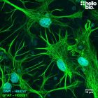
Figure 1. HB6429 staining of NeuN expressing neurons in rat hippocampus.
HB6429 reveals the neuronal architecture of the hippocampal formation. Method: Rat brains were dissected and fixed overnight in 4% PFA before then being incubated in 30% sucrose (in PBS) until sunk (approx. 48hrs). A freezing microtome was used to cut 40µm transverse slices before sections were incubated in 1% NaBH4 for 30 minutes. Sections were blocked in 0.05M glycine, 2% BSA and 3% goat serum before incubation overnight in HB6429 (1:1000 dilution, 1µg/ml). This was followed by a two hour incubation with a polyclonal donkey anti-mouse DyLight 488 conjugated (Thermofisher SA5-10166, 1:300 dilution) secondary antibody. DAPI (HB0747) was used at 1µg/ml to visualise cell nuclei. For more detail please see our IHC(IF) protocol. The image was captured as a tilescan using a Leica DMI6000B inverted epifluorescence microscope coupled to a Photometric Prime 95B camera utilising a 20x objective and DAPI (10ms exposure) / L5 (249ms exposure) filters. Images were captured as a stack (0.98µm z-spacing) before being deconvolved using Huygens Professional software then flattened using a maximum Z projection in ImageJ (Schindelin et al., 2012. Nat Methods, 9(7), 676–682).
Figure 2. NeuN and Tyrosine hydroxylase expression in rat striatum.
HB6429 labels the nuclei of cell bodies in the striatum whereas HB6605 labels dopaminergic projections. Method: Rat brains were dissected and fixed overnight in 4% PFA before then being incubated in 30% sucrose (in PBS) until sunk (approx. 48hrs). A freezing microtome was used to cut 40µm transverse slices before sections were incubated in 1% NaBH4 for 30 minutes. Sections were blocked in 0.05M glycine, 2% BSA and 3% goat serum before incubation overnight in HB6429 (1:1000 dilution, 1µg/ml) and HB6605 (1:1,000 dilution). This was followed by a two hour incubation with polyclonal donkey anti-mouse DyLight 488 conjugated (Thermofisher SA5-10166, 1:300 dilution) and polyclonal goat anti-rabbit Janelia Fluor 525 conjugated secondary antibodies. DAPI (HB0747) was used at 1µg/ml to visualise cell nuclei. For more detail please see our IHC(IF) protocol. The image was captured as using a Leica DMI6000B inverted epifluorescence microscope coupled to a Photometric Prime 95B camera utilising a 10x objective and DAPI (8ms exposure) / L5 (200ms exposure) / RHO (250ms exposure) filters. Images were captured as a stack (4.3µm z-spacing) before being deconvolved using Huygens Professional software then flattened using a maximum Z projection in ImageJ (Schindelin et al., 2012. Nat Methods, 9(7), 676–682).
Figure 3. Independent antibody validation of HB6429 in rat cortex.
HB6429 (mouse monoclonal anti-NeuN) and HB6498 (rabbit polyclonal anti-NeuN) show overlapping patterns of staining in rat cortex providing strong evidence for specificity. Method: Rat brains were dissected and fixed overnight in 4% PFA before then being incubated in 30% sucrose (in PBS) until sunk (approx. 48hrs). A freezing microtome was used to cut 40µm horizontal slices before sections were incubated in 1% NaBH4 for 30 minutes. Sections were blocked in 0.05M glycine, 2% BSA and 3% goat serum before incubation overnight in HB6498 (1:5000 dilution, 0.2µg/ml) and HB6429 (1:1000 dilution, 1µg/ml). This was followed by a two hour incubation with polyclonal goat anti-mouse DyLight 488 conjugated (Thermofisher 35503, 1:300 dilution) and polyclonal goat anti-rabbit DyLight 594 (Thermofisher 35561) secondary antibodies. DAPI (HB0747) was used at 1µg/ml to visualise cell nuclei. For more detail please see our IHC(IF) protocol. The image was captured using a Leica DMI6000B inverted epifluorescence microscope coupled to a Photometric Prime 95B camera utilising a 20x objective and DAP (10ms exposure) / L5 (435ms exposure) / TX2 (118ms exposure) filters. Images were captured as a stack before being deconvolved using Huygens Professional software then flattened using a maximum Z projection in ImageJ (Schindelin et al., 2012. Nat Methods, 9(7), 676–682).
Figure 4. NeuN expression in the rat dentate gyrus visualised using HB6429.
HB6429 successfully stains the dense layer of neurones present in the rat dentate gyrus in PFA fixed sections. Method: hippocampi were dissected from rat brains and fixed overnight in 4% PFA before then being incubated in 30% sucrose (in PBS) for another 24hrs. A freezing microtome was used to cut 40µm transverse slices before sections were incubated in 0.05M glycine for 30 minutes. Sections were blocked in 1% BSA, 22.52mg/ml glycine before incubation overnight in HB6429 (1:500 dilution, 2µg/ml). This was followed by a two hour incubation with secondary antibody (Polyclonal goat anti-mouse DyLight 488 conjugated, Thermofisher 35503, 1:300 dilution). DAPI (HB0747) was used at 1µg/ml to visualise cell nuclei. For more detail please see our IHC(IF) protocol. The image was captured using a Leica DM2500 epifluorescence microscope (10x objective) coupled to a Leica DFC7000T colour digital camera with DAPI (10x gain, 14.4ms exposure) and I3 filters (10x gain, 73.0ms exposure).
Figure 5. NeuN expression in the granule cell layer of the rat dentate gyrus visualised using HB6429.
HB6429 successfully stains the dense layer of neurones present in the rat dentate gyrus in PFA fixed sections. Method: hippocampi were dissected from rat brains and fixed overnight in 4% PFA before then being incubated in 30% sucrose (in PBS) for another 24hrs. A freezing microtome was used to cut 40µm transverse slices before sections were incubated in 0.05M glycine for 30 minutes. Sections were blocked in 1% BSA, 22.52mg/ml glycine before incubation overnight in HB6429 (1:1,000 dilution, 1µg/ml). This was followed by a two hour incubation with secondary antibody (Polyclonal goat anti-mouse DyLight 488 conjugated, Thermofisher 35503, 1:300 dilution). DAPI (HB0747) was used at 1µg/ml to visualise cell nuclei. For more detail please see our IHC(IF) protocol. The image was captured using a Leica SPE confocal laser scanning microscope coupled to a Leica DMi8 inverted epifluorescence microscope. The image was captured using a 40x objective (1x zoom) and 405 (20.0% power, gain: 542) / 488 (27.0% power, gain: 542) laser lines. The image was processed in ImageJ (Schindelin et al., 2012. Nat Methods, 9(7), 676–682).
Figure 6. The effect of varying HB6429 concentration upon staining in rat dentate gyrus
HB6429 successfully labelled the dense layer of neurones present in the granule cell layer of the dentate gyrus in 40µm rat hippocampal sections at a range of dilutions. Method: hippocampi were dissected from rat brains and fixed overnight in 4% PFA before then being incubated in 30% sucrose (in PBS) for another 24hrs. A freezing microtome was used to cut 40µm transverse slices before sections were incubated in 0.05M glycine for 30 minutes. Sections were blocked in 1% BSA, 22.52mg/ml glycine before incubation overnight in varying concentrations of HB6429 (1:500 to 1:4,000 dilutions, 0.25-2µg/ml). This was followed by a two hour incubation with secondary antibody (Polyclonal goat anti-mouse DyLight 488 conjugated, Thermofisher 35503, 1:300 dilution). DAPI (HB0747) was used at 1µg/ml to visualise cell nuclei (not shown due to obscuring NeuN signal in the dense cell layer). For more detail please see our IHC(IF) protocol. Images were captured using a Leica DM2500 epifluorescence microscope (20x objective) coupled to a Leica DFC7000T colour digital camera with DAPI and I3 filters. Exposure times were as follows:
1:500 - I3: 10x gain, 32.2ms exposure
1:1000 - I3: 10x gain, 10x gain, 22.3ms exposure
1:2000 – I3: 10x gain, 22.3ms exposure
1:4000 – I3: 10x gain, 17.9ms exposure
No primary - I3: 112.ms exposure (taken using different microscope: Leica DMI6000B with Photometric-Prime95B camera).
Figure 7. NeuN expression in various tissue lysates and preparations.
HB6429 revealed the double band of FOX-3 isoforms in the brain cytosol fractions and the single heavier band of Synapsin-1 present in P2 brain fractions. These bands are characteristic of antibodies raised against the NeuN antigen. Method: mouse brain and rat brain membrane (P2) and cytosol fractions were prepared following previous work (Molnar et al., 1993. Neuroscience 53:307-326) from freshly collected adult brains. Other tissue lysates were prepared following established protocols from freshly dissected tissue (see our guide on WB sample preparation). Samples were loaded (20µg / lane) onto a 12% acrylamide gel alongside a protein ladder (Thermofisher, 26616) before being run at 60V for 30 minutes followed by 120V for 100 minutes. Wet transfer to a PVDF membrane was completed in 100 minutes using 400mA. The membrane was blocked for 2hrs in 5% non-fat dry milk before being incubated overnight at 4°C in HB6429 at a 1:1,000 dilution (1µg/ml). Following washing, the membrane was incubated in secondary antibody (1:10,000 dilution, Polyclonal goat anti-mouse HRP conjugated, Sigma Aldrich A3682) for 2hrs. For more detail please see our Western blotting protocol. Detection was accomplished using Clarity Western ECL substrate (BioRad, 1705061) and a Licor Odyssey Fc imaging system (ECL channel: 10 min exposure, 700nm channel: 30 sec exposure).
Figure 8. Concentration response of HB6429 staining in rat brain cytosol preparation.
HB6429 shows consistent results with low background at dilutions as low as 1:16,000 (62.5 ng/ml). Method: Rat brain cytosol fractions were prepared following previous work (Molnar et al., 1993. Neuroscience 53:307-326) from freshly collected adult brains. Samples were loaded (20µg / lane) onto a 12% acrylamide gel alongside a protein ladder (Thermofisher, 26616) before being run at 60V for 30 minutes followed by 120V for 100 minutes. Wet transfer to a PVDF membrane was completed in 90 minutes using 400mA. Following transfer the membrane was cut into strips using Ponceau dye to visualise lanes. Strips were blocked for 2hrs in 5% non-fat dry milk before being incubated overnight at 4°C in HB6429. Each strip was incubated separately with a separate HB6429 concentration with this ranging from 2µg/ml (1:500 dilution) to 31.25ng/ml (1:32,000 dilution). Following washing, the membrane was incubated in secondary antibody (1:10,000 dilution, Polyclonal goat anti-mouse HRP conjugated, Sigma Aldrich A3682) for 2hrs. For more detail please see our Western blotting protocol. Detection was accomplished using Clarity Western ECL substrate (BioRad, 1705061) and a Licor Odyssey Fc imaging system (ECL channel: 10 min exposure, 700nm channel: 30 sec exposure). Band intensity was calculated using Image Studio version 5.2.5 (LiCor) and a graph was constructed in GraphPad Prism 9 using a 3-parameter Hill equation curve fit.
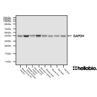
Figure 9. NeuN expression in various tissue lysates and preparations including GAPDH (HB9177) loading control.
HB6429 revealed the complex staining pattern resulting from antibodies raised against the NeuN antigen. Method: mouse brain and rat brain membrane (P2) and cytosol fractions were prepared following previous work (Molnar et al., 1993. Neuroscience 53:307-326) from freshly collected adult brains. Other tissue lysates were prepared following established protocols from freshly dissected tissue (see our guide on WB sample preparation). Samples were loaded (20µg / lane) onto a 12% acrylamide gel alongside a protein ladder (Thermofisher, 26616) before being run at 60V for 35 minutes followed by 120V for 100 minutes. Wet transfer to a PVDF membrane was completed in 90 minutes using 400mA. The membrane was blocked for 2hrs in 5% non-fat dry milk before being incubated overnight at 4°C in HB6429 at a 1:1,000 dilution (1µg/ml) and HB9177 (1:2,000 dilution, 0.5µg/ml). Following washing, the membrane was incubated in secondary antibody (1:10,000 dilution, Polyclonal goat anti-mouse HRP conjugated, Sigma Aldrich A3682) for 2hrs. For more detail please see our Western blotting protocol. Detection was accomplished using Clarity Western ECL substrate (BioRad, 1705061) and a Licor Odyssey Fc imaging system (ECL channel: 10 min exposure, 700nm channel: 30 sec exposure).
Figure 10. NeuN expression in the granule cell layer of the rat dentate gyrus visualised using HB6429.
HB6429 successfully stains the dense layer of neurones present in the rat dentate gyrus in PFA fixed sections. Method: hippocampi were dissected from rat brains and fixed overnight in 4% PFA before then being incubated in 30% sucrose (in PBS) for another 24hrs. A freezing microtome was used to cut 40µm transverse slices before sections were incubated in 0.05M glycine for 30 minutes. Sections were blocked in 1% BSA, 22.52mg/ml glycine before incubation overnight in HB6429 (1:1,000 dilution, 1µg/ml). This was followed by a two hour incubation with secondary antibody (Polyclonal goat anti-mouse DyLight 488 conjugated, Thermofisher 35503, 1:300 dilution). DAPI (HB0747) was used at 1µg/ml to visualise cell nuclei. For more detail please see our IHC(IF) protocol. The image was captured using a Leica SPE confocal laser scanning microscope coupled to a Leica DMi8 inverted epifluorescence microscope. The image was captured using a 40x objective (5.5x zoom) in a z-stack (168nm spacing) and 405 (20.0% power, gain: 494) / 488 (27.0% power, gain: 619) laser lines. The image was processed in ImageJ (Schindelin et al., 2012. Nat Methods, 9(7), 676–682).
Figure 11. NeuN histoblot in a horizontal rat brain section using HB6429.
HB6429 reveals the distribution of NeuN expressing neurons within the rat brain using histoblotting. Note image has been mirrored to show annotated brain regions. Abbreviations: AIV: Agranular insular cortex (ventral), AOP: Anterior olfactory area (posterior), AIP: Agranular insular cortex (posterior), CA1: Hippocampal CA1, CA3: Hippocampal CA3, Cpu: Caudate Putamen, DG: Dentate gyrus, DRN: Dorsal raphe nucleus, ILL: Intermediate nucleus of the lateral lemniscus, Lent: Lateral entorhinal cortex, Nacc: Nucleus accumbens, PB: Parabrachial nucleus, PFl: Paraflocculus PH: Posterior hypothalamic nucleus, PRh: Perirhinal cortex, VN: Vestibularn nucleus. Method: Histoblots were performed following the methodology detailed in Molar, 2016. Neuromethods). In brief: 10µm fresh frozen sections were prepared on a cryostat from rat brains and transferred from slides to nitrocellulose membranes. Membranes were incubated in DNase I for 20 minutes at 37°C before incubation in stripping buffer containing β-mercaptoethanol for 1hr at 45°C. Sections were then blocked in 5% non-fat dry milk in TBST for 1hr before being incubated in HB6429 at 1µg/ml (1:1000 dilution) overnight at 4°C. Following washing, membranes were incubated in secondary antibody (goat anti-mouse alkaline phosphatase conjugated, Thermofisher A16069) at a 1:5,000 dilution for 90 minutes at room temperature. Membranes were developed using HB1881 NBT/BCIP solution before colour development was stopped with PBS. Membranes were imaged on a high-resolution desktop scanner.
Immunostainings comparing NeuN staining in mouse brain slices
Immunostainings comparing NeuN staining in mouse brain slices. The NeuN antibody shows good specificity and signal/noise (S/N). At equivalent dilution, the signal is brighter with this antibody than with our usual antibodies.
Data kindly provided by the Poncer lab, Institute Du Fer À Moulin - Inserm
Figure 1. HB6429 staining of NeuN expressing neurons in rat hippocampus.
HB6429 reveals the neuronal architecture of the hippocampal formation. Method: Rat brains were dissected and fixed overnight in 4% PFA before then being incubated in 30% sucrose (in PBS) until sunk (approx. 48hrs). A freezing microtome was used to cut 40µm transverse slices before sections were incubated in 1% NaBH4 for 30 minutes. Sections were blocked in 0.05M glycine, 2% BSA and 3% goat serum before incubation overnight in HB6429 (1:1000 dilution, 1µg/ml). This was followed by a two hour incubation with a polyclonal donkey anti-mouse DyLight 488 conjugated (Thermofisher SA5-10166, 1:300 dilution) secondary antibody. DAPI (HB0747) was used at 1µg/ml to visualise cell nuclei. For more detail please see our IHC(IF) protocol. The image was captured as a tilescan using a Leica DMI6000B inverted epifluorescence microscope coupled to a Photometric Prime 95B camera utilising a 20x objective and DAPI (10ms exposure) / L5 (249ms exposure) filters. Images were captured as a stack (0.98µm z-spacing) before being deconvolved using Huygens Professional software then flattened using a maximum Z projection in ImageJ (Schindelin et al., 2012. Nat Methods, 9(7), 676–682).
Figure 2. NeuN and Tyrosine hydroxylase expression in rat striatum.
HB6429 labels the nuclei of cell bodies in the striatum whereas HB6605 labels dopaminergic projections. Method: Rat brains were dissected and fixed overnight in 4% PFA before then being incubated in 30% sucrose (in PBS) until sunk (approx. 48hrs). A freezing microtome was used to cut 40µm transverse slices before sections were incubated in 1% NaBH4 for 30 minutes. Sections were blocked in 0.05M glycine, 2% BSA and 3% goat serum before incubation overnight in HB6429 (1:1000 dilution, 1µg/ml) and HB6605 (1:1,000 dilution). This was followed by a two hour incubation with polyclonal donkey anti-mouse DyLight 488 conjugated (Thermofisher SA5-10166, 1:300 dilution) and polyclonal goat anti-rabbit Janelia Fluor 525 conjugated secondary antibodies. DAPI (HB0747) was used at 1µg/ml to visualise cell nuclei. For more detail please see our IHC(IF) protocol. The image was captured as using a Leica DMI6000B inverted epifluorescence microscope coupled to a Photometric Prime 95B camera utilising a 10x objective and DAPI (8ms exposure) / L5 (200ms exposure) / RHO (250ms exposure) filters. Images were captured as a stack (4.3µm z-spacing) before being deconvolved using Huygens Professional software then flattened using a maximum Z projection in ImageJ (Schindelin et al., 2012. Nat Methods, 9(7), 676–682).
Figure 3. Independent antibody validation of HB6429 in rat cortex.
HB6429 (mouse monoclonal anti-NeuN) and HB6498 (rabbit polyclonal anti-NeuN) show overlapping patterns of staining in rat cortex providing strong evidence for specificity. Method: Rat brains were dissected and fixed overnight in 4% PFA before then being incubated in 30% sucrose (in PBS) until sunk (approx. 48hrs). A freezing microtome was used to cut 40µm horizontal slices before sections were incubated in 1% NaBH4 for 30 minutes. Sections were blocked in 0.05M glycine, 2% BSA and 3% goat serum before incubation overnight in HB6498 (1:5000 dilution, 0.2µg/ml) and HB6429 (1:1000 dilution, 1µg/ml). This was followed by a two hour incubation with polyclonal goat anti-mouse DyLight 488 conjugated (Thermofisher 35503, 1:300 dilution) and polyclonal goat anti-rabbit DyLight 594 (Thermofisher 35561) secondary antibodies. DAPI (HB0747) was used at 1µg/ml to visualise cell nuclei. For more detail please see our IHC(IF) protocol. The image was captured using a Leica DMI6000B inverted epifluorescence microscope coupled to a Photometric Prime 95B camera utilising a 20x objective and DAP (10ms exposure) / L5 (435ms exposure) / TX2 (118ms exposure) filters. Images were captured as a stack before being deconvolved using Huygens Professional software then flattened using a maximum Z projection in ImageJ (Schindelin et al., 2012. Nat Methods, 9(7), 676–682).
Figure 4. NeuN expression in the rat dentate gyrus visualised using HB6429.
HB6429 successfully stains the dense layer of neurones present in the rat dentate gyrus in PFA fixed sections. Method: hippocampi were dissected from rat brains and fixed overnight in 4% PFA before then being incubated in 30% sucrose (in PBS) for another 24hrs. A freezing microtome was used to cut 40µm transverse slices before sections were incubated in 0.05M glycine for 30 minutes. Sections were blocked in 1% BSA, 22.52mg/ml glycine before incubation overnight in HB6429 (1:500 dilution, 2µg/ml). This was followed by a two hour incubation with secondary antibody (Polyclonal goat anti-mouse DyLight 488 conjugated, Thermofisher 35503, 1:300 dilution). DAPI (HB0747) was used at 1µg/ml to visualise cell nuclei. For more detail please see our IHC(IF) protocol. The image was captured using a Leica DM2500 epifluorescence microscope (10x objective) coupled to a Leica DFC7000T colour digital camera with DAPI (10x gain, 14.4ms exposure) and I3 filters (10x gain, 73.0ms exposure).
Figure 5. NeuN expression in the granule cell layer of the rat dentate gyrus visualised using HB6429.
HB6429 successfully stains the dense layer of neurones present in the rat dentate gyrus in PFA fixed sections. Method: hippocampi were dissected from rat brains and fixed overnight in 4% PFA before then being incubated in 30% sucrose (in PBS) for another 24hrs. A freezing microtome was used to cut 40µm transverse slices before sections were incubated in 0.05M glycine for 30 minutes. Sections were blocked in 1% BSA, 22.52mg/ml glycine before incubation overnight in HB6429 (1:1,000 dilution, 1µg/ml). This was followed by a two hour incubation with secondary antibody (Polyclonal goat anti-mouse DyLight 488 conjugated, Thermofisher 35503, 1:300 dilution). DAPI (HB0747) was used at 1µg/ml to visualise cell nuclei. For more detail please see our IHC(IF) protocol. The image was captured using a Leica SPE confocal laser scanning microscope coupled to a Leica DMi8 inverted epifluorescence microscope. The image was captured using a 40x objective (1x zoom) and 405 (20.0% power, gain: 542) / 488 (27.0% power, gain: 542) laser lines. The image was processed in ImageJ (Schindelin et al., 2012. Nat Methods, 9(7), 676–682).
Figure 6. The effect of varying HB6429 concentration upon staining in rat dentate gyrus
HB6429 successfully labelled the dense layer of neurones present in the granule cell layer of the dentate gyrus in 40µm rat hippocampal sections at a range of dilutions. Method: hippocampi were dissected from rat brains and fixed overnight in 4% PFA before then being incubated in 30% sucrose (in PBS) for another 24hrs. A freezing microtome was used to cut 40µm transverse slices before sections were incubated in 0.05M glycine for 30 minutes. Sections were blocked in 1% BSA, 22.52mg/ml glycine before incubation overnight in varying concentrations of HB6429 (1:500 to 1:4,000 dilutions, 0.25-2µg/ml). This was followed by a two hour incubation with secondary antibody (Polyclonal goat anti-mouse DyLight 488 conjugated, Thermofisher 35503, 1:300 dilution). DAPI (HB0747) was used at 1µg/ml to visualise cell nuclei (not shown due to obscuring NeuN signal in the dense cell layer). For more detail please see our IHC(IF) protocol. Images were captured using a Leica DM2500 epifluorescence microscope (20x objective) coupled to a Leica DFC7000T colour digital camera with DAPI and I3 filters. Exposure times were as follows:
1:500 - I3: 10x gain, 32.2ms exposure
1:1000 - I3: 10x gain, 10x gain, 22.3ms exposure
1:2000 – I3: 10x gain, 22.3ms exposure
1:4000 – I3: 10x gain, 17.9ms exposure
No primary - I3: 112.ms exposure (taken using different microscope: Leica DMI6000B with Photometric-Prime95B camera).
Figure 7. NeuN expression in various tissue lysates and preparations.
HB6429 revealed the double band of FOX-3 isoforms in the brain cytosol fractions and the single heavier band of Synapsin-1 present in P2 brain fractions. These bands are characteristic of antibodies raised against the NeuN antigen. Method: mouse brain and rat brain membrane (P2) and cytosol fractions were prepared following previous work (Molnar et al., 1993. Neuroscience 53:307-326) from freshly collected adult brains. Other tissue lysates were prepared following established protocols from freshly dissected tissue (see our guide on WB sample preparation). Samples were loaded (20µg / lane) onto a 12% acrylamide gel alongside a protein ladder (Thermofisher, 26616) before being run at 60V for 30 minutes followed by 120V for 100 minutes. Wet transfer to a PVDF membrane was completed in 100 minutes using 400mA. The membrane was blocked for 2hrs in 5% non-fat dry milk before being incubated overnight at 4°C in HB6429 at a 1:1,000 dilution (1µg/ml). Following washing, the membrane was incubated in secondary antibody (1:10,000 dilution, Polyclonal goat anti-mouse HRP conjugated, Sigma Aldrich A3682) for 2hrs. For more detail please see our Western blotting protocol. Detection was accomplished using Clarity Western ECL substrate (BioRad, 1705061) and a Licor Odyssey Fc imaging system (ECL channel: 10 min exposure, 700nm channel: 30 sec exposure).
Figure 8. Concentration response of HB6429 staining in rat brain cytosol preparation.
HB6429 shows consistent results with low background at dilutions as low as 1:16,000 (62.5 ng/ml). Method: Rat brain cytosol fractions were prepared following previous work (Molnar et al., 1993. Neuroscience 53:307-326) from freshly collected adult brains. Samples were loaded (20µg / lane) onto a 12% acrylamide gel alongside a protein ladder (Thermofisher, 26616) before being run at 60V for 30 minutes followed by 120V for 100 minutes. Wet transfer to a PVDF membrane was completed in 90 minutes using 400mA. Following transfer the membrane was cut into strips using Ponceau dye to visualise lanes. Strips were blocked for 2hrs in 5% non-fat dry milk before being incubated overnight at 4°C in HB6429. Each strip was incubated separately with a separate HB6429 concentration with this ranging from 2µg/ml (1:500 dilution) to 31.25ng/ml (1:32,000 dilution). Following washing, the membrane was incubated in secondary antibody (1:10,000 dilution, Polyclonal goat anti-mouse HRP conjugated, Sigma Aldrich A3682) for 2hrs. For more detail please see our Western blotting protocol. Detection was accomplished using Clarity Western ECL substrate (BioRad, 1705061) and a Licor Odyssey Fc imaging system (ECL channel: 10 min exposure, 700nm channel: 30 sec exposure). Band intensity was calculated using Image Studio version 5.2.5 (LiCor) and a graph was constructed in GraphPad Prism 9 using a 3-parameter Hill equation curve fit.
Figure 9. NeuN expression in various tissue lysates and preparations including GAPDH (HB9177) loading control.
HB6429 revealed the complex staining pattern resulting from antibodies raised against the NeuN antigen. Method: mouse brain and rat brain membrane (P2) and cytosol fractions were prepared following previous work (Molnar et al., 1993. Neuroscience 53:307-326) from freshly collected adult brains. Other tissue lysates were prepared following established protocols from freshly dissected tissue (see our guide on WB sample preparation). Samples were loaded (20µg / lane) onto a 12% acrylamide gel alongside a protein ladder (Thermofisher, 26616) before being run at 60V for 35 minutes followed by 120V for 100 minutes. Wet transfer to a PVDF membrane was completed in 90 minutes using 400mA. The membrane was blocked for 2hrs in 5% non-fat dry milk before being incubated overnight at 4°C in HB6429 at a 1:1,000 dilution (1µg/ml) and HB9177 (1:2,000 dilution, 0.5µg/ml). Following washing, the membrane was incubated in secondary antibody (1:10,000 dilution, Polyclonal goat anti-mouse HRP conjugated, Sigma Aldrich A3682) for 2hrs. For more detail please see our Western blotting protocol. Detection was accomplished using Clarity Western ECL substrate (BioRad, 1705061) and a Licor Odyssey Fc imaging system (ECL channel: 10 min exposure, 700nm channel: 30 sec exposure).
Figure 10. NeuN expression in the granule cell layer of the rat dentate gyrus visualised using HB6429.
HB6429 successfully stains the dense layer of neurones present in the rat dentate gyrus in PFA fixed sections. Method: hippocampi were dissected from rat brains and fixed overnight in 4% PFA before then being incubated in 30% sucrose (in PBS) for another 24hrs. A freezing microtome was used to cut 40µm transverse slices before sections were incubated in 0.05M glycine for 30 minutes. Sections were blocked in 1% BSA, 22.52mg/ml glycine before incubation overnight in HB6429 (1:1,000 dilution, 1µg/ml). This was followed by a two hour incubation with secondary antibody (Polyclonal goat anti-mouse DyLight 488 conjugated, Thermofisher 35503, 1:300 dilution). DAPI (HB0747) was used at 1µg/ml to visualise cell nuclei. For more detail please see our IHC(IF) protocol. The image was captured using a Leica SPE confocal laser scanning microscope coupled to a Leica DMi8 inverted epifluorescence microscope. The image was captured using a 40x objective (5.5x zoom) in a z-stack (168nm spacing) and 405 (20.0% power, gain: 494) / 488 (27.0% power, gain: 619) laser lines. The image was processed in ImageJ (Schindelin et al., 2012. Nat Methods, 9(7), 676–682).
Figure 11. NeuN histoblot in a horizontal rat brain section using HB6429.
HB6429 reveals the distribution of NeuN expressing neurons within the rat brain using histoblotting. Note image has been mirrored to show annotated brain regions. Abbreviations: AIV: Agranular insular cortex (ventral), AOP: Anterior olfactory area (posterior), AIP: Agranular insular cortex (posterior), CA1: Hippocampal CA1, CA3: Hippocampal CA3, Cpu: Caudate Putamen, DG: Dentate gyrus, DRN: Dorsal raphe nucleus, ILL: Intermediate nucleus of the lateral lemniscus, Lent: Lateral entorhinal cortex, Nacc: Nucleus accumbens, PB: Parabrachial nucleus, PFl: Paraflocculus PH: Posterior hypothalamic nucleus, PRh: Perirhinal cortex, VN: Vestibularn nucleus. Method: Histoblots were performed following the methodology detailed in Molar, 2016. Neuromethods). In brief: 10µm fresh frozen sections were prepared on a cryostat from rat brains and transferred from slides to nitrocellulose membranes. Membranes were incubated in DNase I for 20 minutes at 37°C before incubation in stripping buffer containing β-mercaptoethanol for 1hr at 45°C. Sections were then blocked in 5% non-fat dry milk in TBST for 1hr before being incubated in HB6429 at 1µg/ml (1:1000 dilution) overnight at 4°C. Following washing, membranes were incubated in secondary antibody (goat anti-mouse alkaline phosphatase conjugated, Thermofisher A16069) at a 1:5,000 dilution for 90 minutes at room temperature. Membranes were developed using HB1881 NBT/BCIP solution before colour development was stopped with PBS. Membranes were imaged on a high-resolution desktop scanner.
Immunostainings comparing NeuN staining in mouse brain slices
Immunostainings comparing NeuN staining in mouse brain slices. The NeuN antibody shows good specificity and signal/noise (S/N). At equivalent dilution, the signal is brighter with this antibody than with our usual antibodies.
Data kindly provided by the Poncer lab, Institute Du Fer À Moulin - Inserm
Product information
Immunogen
Amino acids 213 - 310 of human FOX3 expressed and purified from E. coli
Clone number
1B7
Isotype
IgG2b
Purification
Protein G affinity chromatography
Concentration
1mg/ml
Formulation
50% PBS, 50% glycerol + 5mM sodium azide
Predicted species reactivity
Human, Mouse, Rat
Tested species reactivity
Mouse, Rat
Tested applications
Applications
WB, IHC(IF), Histoblot
Western blot optimal concentration
1:1000 (1μg/ml) as assessed in a rat brain cytosol preparation
IHC(IF) optimal concentration
1:1000 (1μg/ml) as assessed in rat hippocampal sections
ICC optimal concentration
1:1000 (1μg/ml) as assessed in rat horizontal brain sections
Positive control
NeuN is highly expressed in the neurons of the CNS and PNS. It is also expressed in SH-SY5Y cells.
Negative control
Any tissue not of neural origin. Most cell lines are NeuN negative.
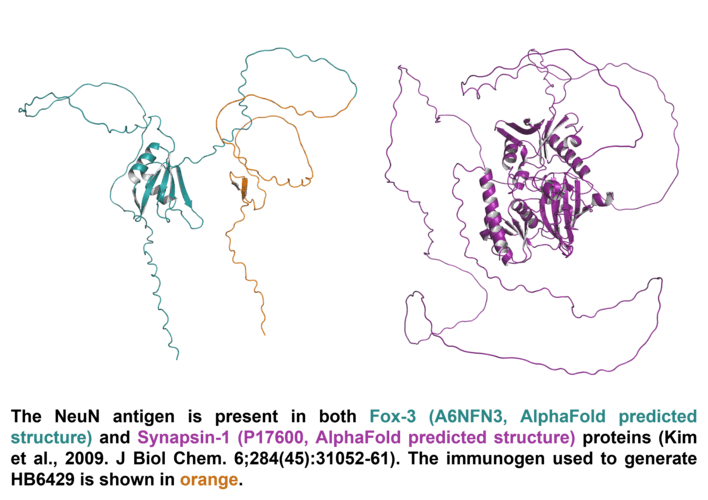
NeuN binds primarily to FOX3 which has two isoforms. Isoform 1 is described as the canonical sequence with 312 amino acids (33.8kDa) while isoform 2 has a 13 residue insert at position 312 leading to a total length of 325 amino acids (35.1kDa). NeuN antibodies also bind to synapsin-1 in western blot experiments (but not in IHC or ICC) which has two isoforms. Isoform 1 is 705aa long (74.1kDa) while isoform 2 is shorter at 669aa (70.0kDa).
Expression
NeuN is expressed only within neurones. While the vast majority of neurones express NeuN some cell types such as Purkinje cells, stellate and golgi cells do not show immunoreactivity.
Subcellular expression
Expression is primarily localised to the nucleus however some FOX3 isoforms can localise to the cytosol.
Target function
FOX3 is a splicing regulator of pre-mRNA responsible for neuronal specific alternative splicing of neuronal proteins.
Processing
None
Post translational modifications
Phosphorylation has been reported (see Lind et al., 2004. J Neurosci Res. 79: 295-302) which is directly related to immunoreactivity whereby dephosphorylation abolished staining.
Homology (compared to human)
Mouse FOX3 shows 95.02% identity to human FOX3 wheras rat FOX3 shows no similarity due to a large 47 residue insertion at amino acid 252 in rats.
Similar proteins
RNA-binding protein fox-1 homolog 1 (40-44kDa) shows 67.3% identity while RNA-binding protein fox-1 homolog 2 (37-47kDa) shows 56.5% identity
Storage & Handling
Storage instructions
-20°C
Shipping Conditions
On ice
Important
This product is for RESEARCH USE ONLY and is not intended for therapeutic or diagnostic use. Not for human or veterinary use
NeuN is a widely used marker for identifying neurons in the central nervous system. It is a nuclear and sometimes perinuclear protein expressed in the majority of post-mitotic (mature) neurons, making it highly specific for neuronal identification.
What mounting media do you recommend to use with this antibody?
We recommend using one of our high performance mounting medias, supplied as either hardset or aqeous with a range of counterstains:
While the vast majority of neurones express NeuN some subtypes such as Purkinje cells, stellate and golgi cells do not show immunoreactivity. NeuN is also not expressed in immature neurones.
What guarantee do you have that my NeuN antibody will perform as expected?
We guarantee that your NeuN antibody will work for the applications and species we list on the datasheet. If the antibody fails to perform as expected then we are happy to offer a 100% refund guarantee. For more details please see our guarantee policy.
Will my NeuN antibody work against species that have not been listed on the datasheet?
A species not being listed doesn’t mean that the antibody won’t work, just that we haven’t tested it. If you test one of our antibodies in a new species please let us know (positive or negative)!
What protocols are available for use with this NeuN antibody
We have made a comprehensive collection of protocols that we have used in our experiments to validate this NeuN antibody.
What counterstains do you recommend for use in ICC and IHC with this NeuN antibody?
We recommend using either DAPI or Hoechst 33342 to label cell nuclei. In some experiments it is also helpful to label actin filaments in the cytoskeleton using a Phalloidin conjugate such as FITC Phalloidin or Rhodamine Phalloidin-TRITC.


 loading control.")


 Western Blot Protocol (1 MB)