

Goat Anti-Rabbit IgG H&L (Biotin) preadsorbed ValidAbTM
Certificate of Analysis
Product overview
| Name | Goat Anti-Rabbit IgG H&L (Biotin) preadsorbed ValidAbTM |
| Host | Goat |
| Clonality | Polyclonal |
| Target | Rabbit IgG H&L |
| Conjugate | Biotin |
| Description | Goat Anti-Rabbit IgG H&L (Biotin) preadrorbed secondary antibody. Part of the ValidAb™ range of highly validated, data-rich antibodies. |
Validation data

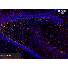
Figure 1. Calretinin positive interneurons and astrocytes in the rat hippocampus.
Method: Rat brains were dissected and fixed overnight in 4% PFA before then being incubated in 30% sucrose (in PBS) until sunk (approx. 48hrs). A freezing microtome was used to cut 40µm horizontal slices before sections were incubated in 1% NaBH4 for 30 minutes. Sections were blocked in 0.05M glycine, 2% BSA and 3% donkey serum before incubation overnight in anti-calretinin HB6494 (1:1,000 dilution) and anti-GFAP HB6406 (1:4000 dilution) at 4°C. This was followed by a two hour incubation with goat anti-rabbit biotin antibody (HB11036) at a dilution of 1:250 and goat anti-chicken Alexa Fluor™ 488 (Invitrogen) at a dilution of 1:300. Following three washes in PBST, sections were incubated with Streptavidin Janelia Fluor® 646 HB17045 at 1.0 µg/mL for 2 hours at room temperature. DAPI HB0747 was used at 1µg/ml to visualise cell nuclei. For more detail please see our IHC(IF) protocol . Images were captured using a Leica SP8 AOBS confocal laser scanning microscope attached to a Leica DM I8 inverted epifluorescence microscope. The image was captured 10x objective, 405nm (6.0% power, PMT: 725.1V gain), 488nm (2.18% power, Hyd: 22.9% gain) and 633nm (10.16% power, Hyd: 94.7% gain) laser lines in a z-stack (3.67µm spacing). The stack was flattened using a maximum Z projection in ImageJ (Schindelin et al., 2012. Nat Methods, 9(7), 676–682).

Figure 2. Tyrosine hydroxylase staining in rat striatum visualized using biotin-streptavidin detection.

Figure 3. Calretinin and Parvalbumin expression in rat hippocampus.
Method: Rat brains were dissected and fixed overnight in 4% PFA before incubation in 30% sucrose (in PBS) until sunk (approx. 48hrs). A freezing microtome was used to cut 40µm horizontal slices before sections were incubated in 1% NaBH4 for 30 minutes. Sections were blocked in 0.05M glycine, 2% BSA and 3% donkey serum before incubation overnight in anti-calretinin HB6494 (1:4,000 dilution) and anti-parvalbumin HB6457 (1:1000 dilution) at 4°C. This was followed by a two hour incubation with goat anti-rabbit biotin antibody (HB11036) at a dilution of 1:250, and goat anti-mouse Dylight™ 594 (Thermofisher) at a dilution of 1:300. Following three washes in PBST, sections were incubated with Streptavidin Janelia Fluor® 646 HB6494 at 1.0 µg/mL for 2 hours at room temperature. DAPI HB0747 was used at 1µg/ml to visualise cell nuclei. For more detail please see our IHC(IF) protocol. Images were captured using a Leica SP8 AOBS confocal laser scanning microscope attached to a Leica DM I8 inverted epifluorescence microscope. The image was captured using a 10x objective, 405nm (6.0% power, PMT: 725.1V gain), 580nm (3.63% power, Hyd: 68% gain) and 633nm (10.16% power, Hyd: 94.7% gain) laser lines in a z-stack (3.67µm spacing). The stack was flattened using a maximum Z projection in ImageJ (Schindelin et al., 2012. Nat Methods, 9(7), 676–682).

Figure 4. Tyrosine hydroxylase staining in rat striatum visualized using biotin-streptavidin detection.

Figure 5. Concentration response of HB17046 staining in rat hippocampus.
Method: Rat brains were dissected and fixed overnight in 4% PFA before then being incubated in 30% sucrose (in PBS) until sunk (approx. 48hrs). A freezing microtome was used to cut 40µm horizontal slices before sections were incubated in 1% NaBH4 for 30 minutes. Sections were blocked in 0.05M glycine, 2% BSA and 3% donkey serum before incubation overnight in anti-parvalbumin HB6494 (1:4,000 dilution) at 4°C. This was followed by a two hour incubation with goat anti-rabbit biotin antibody HB11345 at a dilution of 1:250. Following three washes in PBST, sections were incubated with Streptavidin Janelia Fluor® 646 HB17045) at 0.5 µg/mL, 1.0 µg/mL or 5.0 µg/mL for 2 hours at room temperature. DAPI HB0747 was used at 1µg/ml to visualise cell nuclei. For more detail please see our IHC(IF) protocol . Images were captured using a Leica DMI6000B inverted epifluorescence microscope. Images were captured using a 20x objective in a z-stack with exposures:
- 0.5 µg/mL: DAP: 46.156 ms, Y5: 11222.92 ms
- 1.0 µg/mL: DAP: 46.156 ms, Y5: 6131.079 ms
- 5.0 µg/mL: DAP: .756 ms, Y5: 11222.92 ms
Stacks were deconvolved using Huygens professional then flattened using a maximum Z projection in ImageJ (Schindelin et al., 2012. Nat Methods, 9(7), 676–682).

Figure 6. Tyrosine hydroxylase staining in rat striatum visualized using biotin-streptavidin detection.
Figure 1. Calretinin positive interneurons and astrocytes in the rat hippocampus.
Method: Rat brains were dissected and fixed overnight in 4% PFA before then being incubated in 30% sucrose (in PBS) until sunk (approx. 48hrs). A freezing microtome was used to cut 40µm horizontal slices before sections were incubated in 1% NaBH4 for 30 minutes. Sections were blocked in 0.05M glycine, 2% BSA and 3% donkey serum before incubation overnight in anti-calretinin HB6494 (1:1,000 dilution) and anti-GFAP HB6406 (1:4000 dilution) at 4°C. This was followed by a two hour incubation with goat anti-rabbit biotin antibody (HB11036) at a dilution of 1:250 and goat anti-chicken Alexa Fluor™ 488 (Invitrogen) at a dilution of 1:300. Following three washes in PBST, sections were incubated with Streptavidin Janelia Fluor® 646 HB17045 at 1.0 µg/mL for 2 hours at room temperature. DAPI HB0747 was used at 1µg/ml to visualise cell nuclei. For more detail please see our IHC(IF) protocol . Images were captured using a Leica SP8 AOBS confocal laser scanning microscope attached to a Leica DM I8 inverted epifluorescence microscope. The image was captured 10x objective, 405nm (6.0% power, PMT: 725.1V gain), 488nm (2.18% power, Hyd: 22.9% gain) and 633nm (10.16% power, Hyd: 94.7% gain) laser lines in a z-stack (3.67µm spacing). The stack was flattened using a maximum Z projection in ImageJ (Schindelin et al., 2012. Nat Methods, 9(7), 676–682).
Figure 2. Tyrosine hydroxylase staining in rat striatum visualized using biotin-streptavidin detection.
Figure 3. Calretinin and Parvalbumin expression in rat hippocampus.
Method: Rat brains were dissected and fixed overnight in 4% PFA before incubation in 30% sucrose (in PBS) until sunk (approx. 48hrs). A freezing microtome was used to cut 40µm horizontal slices before sections were incubated in 1% NaBH4 for 30 minutes. Sections were blocked in 0.05M glycine, 2% BSA and 3% donkey serum before incubation overnight in anti-calretinin HB6494 (1:4,000 dilution) and anti-parvalbumin HB6457 (1:1000 dilution) at 4°C. This was followed by a two hour incubation with goat anti-rabbit biotin antibody (HB11036) at a dilution of 1:250, and goat anti-mouse Dylight™ 594 (Thermofisher) at a dilution of 1:300. Following three washes in PBST, sections were incubated with Streptavidin Janelia Fluor® 646 HB6494 at 1.0 µg/mL for 2 hours at room temperature. DAPI HB0747 was used at 1µg/ml to visualise cell nuclei. For more detail please see our IHC(IF) protocol. Images were captured using a Leica SP8 AOBS confocal laser scanning microscope attached to a Leica DM I8 inverted epifluorescence microscope. The image was captured using a 10x objective, 405nm (6.0% power, PMT: 725.1V gain), 580nm (3.63% power, Hyd: 68% gain) and 633nm (10.16% power, Hyd: 94.7% gain) laser lines in a z-stack (3.67µm spacing). The stack was flattened using a maximum Z projection in ImageJ (Schindelin et al., 2012. Nat Methods, 9(7), 676–682).
Figure 4. Tyrosine hydroxylase staining in rat striatum visualized using biotin-streptavidin detection.
Figure 5. Concentration response of HB17046 staining in rat hippocampus.
Method: Rat brains were dissected and fixed overnight in 4% PFA before then being incubated in 30% sucrose (in PBS) until sunk (approx. 48hrs). A freezing microtome was used to cut 40µm horizontal slices before sections were incubated in 1% NaBH4 for 30 minutes. Sections were blocked in 0.05M glycine, 2% BSA and 3% donkey serum before incubation overnight in anti-parvalbumin HB6494 (1:4,000 dilution) at 4°C. This was followed by a two hour incubation with goat anti-rabbit biotin antibody HB11345 at a dilution of 1:250. Following three washes in PBST, sections were incubated with Streptavidin Janelia Fluor® 646 HB17045) at 0.5 µg/mL, 1.0 µg/mL or 5.0 µg/mL for 2 hours at room temperature. DAPI HB0747 was used at 1µg/ml to visualise cell nuclei. For more detail please see our IHC(IF) protocol . Images were captured using a Leica DMI6000B inverted epifluorescence microscope. Images were captured using a 20x objective in a z-stack with exposures:
- 0.5 µg/mL: DAP: 46.156 ms, Y5: 11222.92 ms
- 1.0 µg/mL: DAP: 46.156 ms, Y5: 6131.079 ms
- 5.0 µg/mL: DAP: .756 ms, Y5: 11222.92 ms
Stacks were deconvolved using Huygens professional then flattened using a maximum Z projection in ImageJ (Schindelin et al., 2012. Nat Methods, 9(7), 676–682).
Figure 6. Tyrosine hydroxylase staining in rat striatum visualized using biotin-streptavidin detection.
Product information
| Immunogen | Purified rabbit IgG |
| Isotype | IgG |
| Purification | Immunogen affinity chromatography. Pre-adsorbed with mouse and human serum proteins |
| Concentration | 0.5 mg/ml |
| Formulation | Lyophilised. When reconstituted contains PBS with 1% recombinant albumin and less than 0.1% sodium azide |
Tested applications
| Applications | ELISA, ICC, IHC, IHC-P, WB, IHC(IF) |
| Western blot optimal concentration | Tested at 1:10,000. Optimise dependent upon assay. |
| IHC(IF) optimal concentration | Tested at 1:250 dilution (2µg/ml). Optimise dependent upon assay. |
| IHC-P optimal concentration | Tested at 1:300 dilution. Optimise dependent upon assay. |
| ICC optimal concentration | Tested at 1:250 dilution (2µg/ml). Optimise dependent upon assay. |
| Negative control | While this antibody has been cross-adsorbed to reduce non-specific binding it is still often worthwhile to conduct a control experiment where the primary antibody is omitted to give confidence that the staining pattern observed is specific. |
Storage & Handling
| Storage instructions | -20°C then use reconstitution advice |
| Reconstitution advice | We recommend reconstituting with either:
Take care when opening as the precipitate is extremely light and can easily be lost if disturbed. When reconstituting make sure that the antibody is thoroughly dissolved by pipetting up and down before giving the antibody a brief spin at <10,000g to make sure that all material is recovered and at the bottom of the tube. For more information please see our detailed guide on storing and using your antibody |
| Shipping Conditions | Stable for ambient temperature shipping. Follow storage instructions on receipt. |
| Important | This product is for RESEARCH USE ONLY and is not intended for therapeutic or diagnostic use. Not for human or veterinary use |
Technical guides
 Western Blot Protocol (1 MB) Immunocytochemistry Protocol (1.2 MB) Immunohistochemistry Protocol (1.6 MB)
 References for Goat Anti-Rabbit IgG H&L (Biotin) preadsorbed ValidAbTM
-
Antigen Retrieval and Signal Amplification.
Dunkenberger L et al (2022) Methods in molecular biology (Clifton, N.J.) 2422 : 65-74 -
The use of antiavidin antibody and avidin-biotin-peroxidase complex in immunoperoxidase technics.
Hsu SM et al (1981) American journal of clinical pathology 75 : 816-21

Related Products
- Code:
- HB11345
Goat Anti-Mouse IgG H&L (Biotin) preadsorbed secondary antibody. Part of the ValidAb™ range of highly validated, data-rich antibodies.