Anti-c-Fos antibody ValidAbTM

(HB8006)
Technical documents: SDS Datasheet

Product overview

Name Anti-c-Fos antibody ValidAbTM
Host Mouse
Clonality Monoclonal
Target c-Fos
Description

Antibody to c-Fos - an immediate early gene used as a marker of neuronal activity. Part of the ValidAb™ range of highly validated, data-rich antibodies.

Write Your Own Review
You're reviewing:Anti-c-Fos antibody ValidAb<sup>TM</sup>
Rate this item:

Validation data

Figure 1. c-Fos expression in the dentate gyrus of TRAP2;Ai14 mice.

HB8006 reveals c-Fos positive neurons activated during re-exposure to a learning event alongside tdTomato expression in cells activated during the initial learning event. Method: TRAP2;Ai14 mice were administered tamoxifen and underwent a learning experience (driving expression of tdTomato in activated neurones). Seven days later animals were re-exposed to the stimulus then trans-cardiac perfused 90 minutes after behaviour with 0.1M PB (pH7.4) followed by 4% PFA. Brains were extracted and then post-fixed in 4% PFA for 2 hours followed by incubation in 30% sucrose until the brain sank. Coronal sections were cut on a cryostat and stored in cryopreserve at -20°C before further processing. Sections were washed in PBS before being incubated for 30 minutes in sodium citrate buffer (pH 6.0) at 80°C. After allowing sections to return to ambient temperature they were washed in PBST (PBS, 0.2% Triton X-100) before being incubated in mouse on mouse blocking buffer for 1 hour (2 drops per 2.5ml PBST, Vector laboratories, MKB-2213-1). Sections were then incubated in 5% goat serum, 2.5% BSA (blocking buffer) for 1 hour before being incubated for 48hrs at 4°C in HB8006 (1:1000 dilution in blocking buffer, 1µg/ml). Following washing, sections were then incubated in secondary antibody (Goat anti-mouse Alexa Fluor 488 conjugated, 1:500 dilution in blocking buffer, Invitrogen, A-11029) for 24hrs at 4°C. Following washing, sections were mounted using Vectashield Hardset with DAPI (H-1500) and imaged using a 100x objective using a epifluorescence microscope. Data was kindly provided by Gareth Barker at the University of Bristol.

Figure 2. c-Fos expression in the cortex of TRAP2;Ai14 mice.

HB8006 reveals c-Fos positive neurons activated during re-exposure to a learning event alongside tdTomato expression in cells activated during the initial learning event. Method: TRAP2;Ai14 mice were administered tamoxifen and underwent a learning experience (driving expression of tdTomato in activated neurones). Seven days later animals were re-exposed to the stimulus then trans-cardiac perfused 90 minutes after behaviour with 0.1M PB (pH7.4) followed by 4% PFA. Brains were extracted and then post-fixed in 4% PFA for 2 hours followed by incubation in 30% sucrose until the brain sank. Coronal sections were cut on a cryostat and stored in cryopreserve at -20°C before further processing. Sections were washed in PBS before being incubated for 30 minutes in sodium citrate buffer (pH 6.0) at 80°C. After allowing sections to return to ambient temperature they were washed in PBST (PBS, 0.2% Triton X-100) before being incubated in mouse on mouse blocking buffer for 1 hour (2 drops per 2.5ml PBST, Vector laboratories, MKB-2213-1). Sections were then incubated in 5% goat serum, 2.5% BSA (blocking buffer) for 1 hour before being incubated for 48hrs at 4°C in HB8006 (1:1000 dilution in blocking buffer, 1µg/ml). Following washing, sections were then incubated in secondary antibody (Goat anti-mouse Alexa Fluor 488 conjugated, 1:500 dilution in blocking buffer, Invitrogen, A-11029) for 24hrs at 4°C. Following washing, sections were mounted using Vectashield Hardset with DAPI (H-1500) and imaged using a 100x objective using a epifluorescence microscope. Data was kindly provided by Gareth Barker at the University of Bristol.

Figure 3. c-Fos expression in mouse dentate gyrus.

HB8006 reveals c-Fos positive neurons activated during re-exposure to a learning event. Method: TRAP2;Ai14 mice underwent an initial learning experience followed by re-exposure 7 days later. 90 minutes after behaviour, animals were trans-cardiac perfused with 0.1M PB (pH7.4) followed by 4% PFA. Brains were extracted and then post-fixed in 4% PFA for 2 hours followed by incubation in 30% sucrose until the brain sank. Coronal sections were cut on a cryostat and stored in cryopreserve at -20°C before further processing. Sections were washed in PBS before being incubated for 30 minutes in sodium citrate buffer (pH 6.0) at 80°C. After allowing sections to return to ambient temperature they were washed in PBST (PBS, 0.2% Triton X-100) before being incubated in mouse on mouse blocking buffer for 1 hour (2 drops per 2.5ml PBST, Vector laboratories, MKB-2213-1). Sections were then incubated in 5% goat serum, 2.5% BSA (blocking buffer) for 1 hour before being incubated for 48hrs at 4°C in HB8006 (1:1000 dilution in blocking buffer, 1µg/ml). Following washing, sections were then incubated in secondary antibody (Goat anti-mouse Alexa Fluor 488 conjugated, 1:500 dilution in blocking buffer, Invitrogen, A-11029) for 24hrs at 4°C. Following washing, sections were mounted using Vectashield Hardset with DAPI (H-1500) and imaged using a 100x objective using a epifluorescence microscope. Data was kindly provided by Gareth Barker at the University of Bristol.

Product information

Immunogen

Full length recombinant human c-Fos expressed in and purified from E. coli.

Clone number 2H2
Isotype IgG1
Purification

Protein G affinity chromatography

Concentration 1 mg/ml
Formulation 50% PBS, 50% glycerol with 5mM sodium azide
Predicted species reactivity Mouse, Rat, Human
Tested species reactivity Mouse

Tested applications

Applications IHC(IF)
IHC(IF) optimal concentration

1μg/ml (1:1000 dilution) as tested in 4% PFA fixed mouse brain tissue

Product specific protocols
  • Due to the low stability of the c-Fos protein (around a 1hr half life) we recommend perfusion fixation for animal tissues to ensure rapid preservation of protein integrity. A protocol for this is available in Gage et al., 2012.
  • This product has only been validated in IHC(IF) using sodium citrate antigen retrieval (pH6.0, 80°C for 30 minutes) and therefore it is highly recommended that this method is used in all IHC(IF) experiments using HB8006.
Positive control

c-Fos is expressed at high levels in many cell lines (e.g. HEK293 or HeLa) when serum starved cells are stimulated with serum. 

Negative control

Serum starved cell lines (e.g. HEK293 or HeLa) express very low levels of c-Fos. 

Open data link

Please follow this link to the OSF

Target information

Other names
  • Fos 
  • Cellular oncogene fos 
  • Fos proto-oncogene, AP-1 transcription factor subunit 
  • G0/G1 switch regulatory protein 7 
  • Proto-oncogene c-Fos 
  • Transcription factor AP-1 subunit c-Fos 
UniProt ID P01100
Structure image  Chemical Structure
Gene name FOS
NCBI full gene name Fos proto-oncogene, AP-1 transcription factor subunit
Entrez gene ID

2353

Amino acids

380aa (40.7kDa)

Isoforms

c-Fos has three key isoforms:

  • Isoform 1: canonical 380aa, 40.7kDa 
  • Isoform 2: missing aa1-114, 266aa, 28.9kDa 
  • Isoform 3: missing aa132-167, 344aa, 36.3kDa 
Expression

Expressed widely across multiple tissues at low levels. Expression is inducible by a range of factors including cellular activity, growth factors, cytokines and tumour promoters. c-Fos is expressed in neurones where its expression is induced by activity.

Subcellular expression

c-Fos expression is localised to the nucleus 

Processing

None

Post translational modifications

Subject to phosphorylation on multiple residues and also possesses multiple SUMO2 binding sites. 

Homology (compared to human)

Rat and mouse show a 94.2% and 93.7% identity to human c-Fos in a BLAST search

Similar proteins

The following proteins were identified as being similar to c-Fos in a BLAST search:

  • FosB - 73.9% identity 
  • FRA-1 - 52.7% identity 
  • FRA-2 - 42.9% identity 

Storage & Handling

Storage instructions

-20°C

Important This product is for RESEARCH USE ONLY and is not intended for therapeutic or diagnostic use. Not for human or veterinary use

FAQs

What counterstains do you recommend for use in ICC and IHC with this c-Fos antibody?

We recommend using either DAPI, Hoechst 33342 or Propidium Iodide to label cell nuclei. In some experiments it is also helpful to label actin filaments in the cytoskeleton using a Phalloidin conjugate such as FITC Phalloidin or Rhodamine Phalloidin-TRITC.

Will my c-Fos antibody work against species that have not been listed on the datasheet?

A species not being listed doesn’t mean that the c-Fos antibody won’t work, just that we haven’t tested it. If you test one of our antibodies in a new species please let us know (positive or negative)!

What protocols are available for use with this c-Fos antibody?

We have made a comprehensive collection of protocols that we have used in our experiments to validate this c-Fos antibody.

What guarantee do you have that my c-Fos antibody will perform as expected?

We guarantee that your c-Fos antibody will work for the applications and species we list on the datasheet. If the antibody fails to perform as expected then we are happy to offer a 100% refund guarantee. For more details please see our guarantee policy.
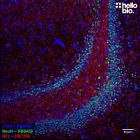
What mounting media do you recommend to use with this antibody?
What other neuroscience markers are available?
Any other questions?

For any other questions about our antibody products please see our technical FAQs for antibodies

References for Anti-c-Fos antibody ValidAbTM

References are publications that support the biological activity of the product
  • Expression of c-fos-like protein as a marker for neuronal activity following noxious stimulation in the rat.

    Bullitt E (1990) The Journal of comparative neurology 296 : 517-30