Antibody to tyrosine hydroxylase (TH) - the rate limiting enzyme in catecholamine synthesis and used as a marker for catecholaminergic (dopaminergic and noradrenergic) neurones in the CNS. Part of the ValidAb™ range of highly validated, data-rich antibodies.
Midbrain dopaminergic neurones stained for tyrosine hydroxylase with HB6589. Method: Rat brains were dissected and fixed overnight in 4% PFA before then being incubated in 30% sucrose (in PBS) until sunk (approx. 48hrs). A freezing microtome was used to cut 40µm horizontal slices before sections were incubated in 1% NaBH4 for 15 minutes. Sections were blocked in 0.05M glycine, 2% BSA and 3% goat serum before incubation overnight in HB6589 (1:1000 dilution). This was followed by a two-hour incubation with secondary antibody at a 1:300 dilution (polyclonal donkey anti-chicken DyLight 488, Thermofisher, A78948). DAPI (HB0747) was used at 1µg/ml to visualise cell nuclei and sections were mounted using MightyMount Antifade Fluorescence Mounting Medium (hardset). For more detail please see our IHC(IF) protocol. Images were captured using a Leica DMI6000B inverted epifluorescence microscope. The image was captured using a 100x objective in a z-stack (0.22µm spacing). The image was captured as a tilescan using A4 (72ms @1x gain exposure) and GFP (70ms @ 1x gain exposure) filters. The stack was deconvolved using Huygens professional then flattened using a maximum Z projection in ImageJ (Schindelin et al., 2012. Nat Methods, 9(7), 676–682).
Figure 2. Rat striatum stained for tyrosine hydroxylase and myelin basic protein with HB6589 and HB8014.
Rat striatum showing dopaminergic terminals stained for tyrosine hydroxylase with HB6589 and myelin stained for myelin basic protein (MBP) with HB8014. Method: Rat brains were dissected and fixed overnight in 4% PFA before then being incubated in 30% sucrose (in PBS) until sunk (approx. 48hrs). A freezing microtome was used to cut 40µm horizontal slices before sections were incubated in 1% NaBH4 for 15 minutes. Sections were blocked in 0.05M glycine, 2% BSA and 3% goat serum before incubation overnight in HB6589 (1:5,000 dilution) and HB8014 (1:1,000 dilution). This was followed by a two-hour incubation with secondary antibodies at a 1:300 dilution (polyclonal donkey anti-chicken DyLight 488, Thermofisher, A78948 and polyclonal goat anti-mouse DyLight 550, Thermofisher, 84540). Sections were mounted using MightyMount Antifade Fluorescence Mounting Medium with DAPI (aqueous) For more detail please see our IHC(IF) protocol. For more detail please see our IHC(IF) protocol. Images were captured using a Leica DMI6000B inverted epifluorescence microscope. The image was captured using a 20x objective in a z-stack (0.59µm spacing). The image was captured as a tilescan using DAP (20ms exposure), L5 (150ms exposure) and TX2 (22.8ms exposure) filters. The stack was deconvolved using Huygens professional then flattened using a maximum Z projection in ImageJ (Schindelin et al., 2012. Nat Methods, 9(7), 676–682).
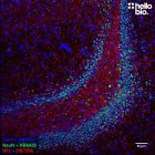
Figure 3. Independent antibody validation of HB6589 and HB6605 in rat CPu.
The staining pattens of HB6589 and HB6605 overlap in rat caudate putamen (CPu) showing strong evidence for antibody specificity. Method: Rat brains were dissected and fixed overnight in 4% PFA before then being incubated in 30% sucrose (in PBS) until sunk (approx. 48hrs). A freezing microtome was used to cut 40µm horizontal slices before sections were incubated in 1% NaBH4 for 15 minutes. Sections were blocked in 0.05M glycine, 2% BSA and 3% goat serum before incubation overnight in HB6589 (1:5,000 dilution), HB8014 (1:1,000 dilution) and HB6605 (1:4,000 dilution). This was followed by a two-hour incubation with secondary antibodies at a 1:300 dilution (polyclonal donkey anti-chicken DyLight 488, Thermofisher, A78948, polyclonal goat anti-mouse DyLight 550, Thermofisher, 84540 and polyclonal goat anti-rabbit DyLight 650, Thermofisher, 11804574). Sections were mounted using MightyMount Antifade Fluorescence Mounting Medium with DAPI (aqueous). For more detail please see our IHC(IF) protocol. Images were captured using a Leica DMI6000B inverted epifluorescence microscope. The image was captured using a 10x objective in a z-stack (5µm spacing). The image was captured as a tilescan using DAP (38ms exposure), L5 (264ms exposure), TX2 (61.8ms exposure) and Y5 (500ms exposure) filters. The stack was deconvolved using Huygens professional then flattened using a maximum Z projection in ImageJ (Schindelin et al., 2012. Nat Methods, 9(7), 676–682).
Figure 4. Concentration response of HB6589 staining in rat striatum.
HB6589 stains rat dopaminergic terminals in the striatum with high signal to noise at dilutions down to 1:8,000. Method: Rat brains were dissected and fixed overnight in 4% PFA before then being incubated in 30% sucrose (in PBS) until sunk (approx. 48hrs). A freezing microtome was used to cut 40µm horizontal slices before sections were incubated in 1% NaBH4 for 15 minutes. Sections were blocked in 0.05M glycine, 2% BSA and 3% goat serum before incubation overnight in HB7167 (1:1,000 (1µg/ml) to 1:8,000 (0.125µg/ml) dilutions). This was followed by a two-hour incubation with secondary antibody at a 1:300 dilution (polyclonal donkey anti-chicken DyLight 488, Thermofisher, A78948). DAPI (HB0747) was used at 1µg/ml to visualise cell nuclei and sections were mounted using MightyMount Antifade Fluorescence Mounting Medium (hardset). For more detail please see our IHC(IF) protocol. Images were captured using a Leica DMI6000B inverted epifluorescence microscope. Images were captured using a 20x objective in a z-stack (0.59µm spacing). Stacks were deconvolved using Huygens professional then flattened using a maximum Z projection in ImageJ (Schindelin et al., 2012. Nat Methods, 9(7), 676–682). Exposure times were as follows:
• 1:1,000 – DAP: 24.5ms, L5: 44.8ms
• 1:2,000 – DAP: 20ms, L5: 113.2ms
• 1:4,000 – DAP: 25ms, L5: 153.8ms
• 1:8,000 – DAP: 20ms, L5: 267.4ms
Figure 5. MBP and Tyrosine hydroxylase staining in rat striatum
Rat striatum showing dopaminergic terminals stained for tyrosine hydroxylase with HB6589 and myelin stained for myelin basic protein (MBP) with HB8014. Method: Rat brains were dissected and fixed overnight in 4% PFA before then being incubated in 30% sucrose (in PBS) until sunk (approx. 48hrs). A freezing microtome was used to cut 40µm horizontal slices before sections were incubated in 1% NaBH4 for 15 minutes. Sections were blocked in 0.05M glycine, 2% BSA and 3% goat serum before incubation overnight in HB6589 (1:5,000 dilution) and HB8014 (1:1,000 dilution). This was followed by a two-hour incubation with secondary antibodies at a 1:300 dilution (polyclonal donkey anti-chicken DyLight 488, Thermofisher, A78948 and polyclonal goat anti-mouse DyLight 550, Thermofisher, 84540). Sections were mounted using MightyMount Antifade Fluorescence Mounting Medium with DAPI (aqueous). For more detail please see our IHC(IF) protocol. Images were captured using a Leica DMI6000B inverted epifluorescence microscope. The image was captured using a 40x objective in a z-stack (0.55µm spacing). The image was captured as a tilescan using DAP (16ms exposure), L5 (76ms exposure) and TX2 (15ms exposure) filters. The stack was deconvolved using Huygens professional then flattened using a maximum Z projection in ImageJ (Schindelin et al., 2012. Nat Methods, 9(7), 676–682).
Figure 6. βIII tubulin and tyrosine hydroxylase staining in rat caudate putamen.
Rat caudate putamen showing dopaminergic terminals stained for tyrosine hydroxylase with HB6589 and neurons stained for βIII-tubulin with HB6639. Method: Rat brains were dissected and fixed overnight in 4% PFA before then being incubated in 30% sucrose (in PBS) until sunk (approx. 48hrs). A freezing microtome was used to cut 40µm horizontal slices before sections were incubated in 1% NaBH4 for 15 minutes. Sections were blocked in 0.05M glycine, 2% BSA and 3% goat serum before incubation overnight in HB6589 (1:2,000 dilution) and HB6639 (1:2,000 dilution). This was followed by a two-hour incubation with secondary antibodies at a 1:300 dilution (polyclonal donkey anti-chicken DyLight 488, Thermofisher, A78948 and polyclonal goat anti-mouse DyLight 550, Thermofisher, 84540). DAPI (HB0747) was used at 1µg/ml to visualise cell nuclei and sections were mounted using MightyMount Antifade Fluorescence Mounting Medium (hardset). For more detail please see our IHC(IF) protocol. The image was captured using a Leica DMI6000B inverted epifluorescence microscope. The image was captured using a 20x objective in a z-stack (0.59µm spacing). The image was captured as a tilescan using DAP (20ms exposure), L5 (113ms exposure) and TX2 (176ms exposure) filters. The stack was deconvolved using Huygens professional then flattened using a maximum Z projection in ImageJ (Schindelin et al., 2012. Nat Methods, 9(7), 676–682).
Figure 7. Independent antibody validation of HB7167 and HB6589 in midbrain dopaminergic neurons.
The staining patterns of HB7176 and HB6589 completely overlap in midbrain dopaminergic neurons showing strong evidence for antibody specificity. Method: Rat brains were dissected and fixed overnight in 4% PFA before then being incubated in 30% sucrose (in PBS) until sunk (approx. 48hrs). A freezing microtome was used to cut 40µm horizontal slices before sections were incubated in 1% NaBH4 for 15 minutes. Sections were blocked in 0.05M glycine, 2% BSA and 3% goat serum before incubation overnight in HB7176 (1:1,000 dilution) and HB6589 (1:1,000 dilution). This was followed by a two-hour incubation with secondary antibodies at a 1:300 dilution (polyclonal goat anti-mouse DyLight 550, Thermofisher, 84540 and polyclonal donkey anti-chicken DyLight 488, Thermofisher, A78948). DAPI (HB0747) was used at 1µg/ml to visualise cell nuclei and sections were mounted using MightyMount Antifade Fluorescence Mounting Medium (hardset). For more detail please see our IHC(IF) protocol. The image was captured using a Leica DMI6000B inverted epifluorescence microscope. The image was captured using a 40x objective in a z-stack (0.3µm spacing). The image was captured as a tilescan using DAP (30ms exposure), L5 (7ms exposure) and RHO (5ms exposure) filters. The stack was deconvolved using Huygens professional then flattened using a maximum Z projection in ImageJ (Schindelin et al., 2012. Nat Methods, 9(7), 676–682).
Figure 8. Independent antibody validation of HB7167 and HB6589 in dopaminergic projections.
The staining patterns of HB7176 and HB6589 completely overlap in rat dopaminergic projections showing strong evidence for antibody specificity. Method: Rat brains were dissected and fixed overnight in 4% PFA before then being incubated in 30% sucrose (in PBS) until sunk (approx. 48hrs). A freezing microtome was used to cut 40µm horizontal slices before sections were incubated in 1% NaBH4 for 15 minutes. Sections were blocked in 0.05M glycine, 2% BSA and 3% goat serum before incubation overnight in HB7176 (1:1,000 dilution) and HB6589 (1:1,000 dilution). This was followed by a two-hour incubation with secondary antibodies at a 1:300 dilution (polyclonal goat anti-mouse DyLight 550, Thermofisher, 84540 and polyclonal donkey anti-chicken DyLight 488, Thermofisher, A78948). DAPI (HB0747) was used at 1µg/ml to visualise cell nuclei and sections were mounted using MightyMount Antifade Fluorescence Mounting Medium (hardset). For more detail please see our IHC(IF) protocol. The image was captured using a Leica DMI6000B inverted epifluorescence microscope. The image was captured using a 40x objective in a z-stack (0.62µm spacing). The image was captured as a tilescan using DAP (49ms exposure), L5 (15ms exposure) and RHO (10ms exposure) filters. The stack was deconvolved using Huygens professional then flattened using a maximum Z projection in ImageJ (Schindelin et al., 2012. Nat Methods, 9(7), 676–682).
Figure 9. Independent antibody validation of HB6589 in rat caudate putamen.
The staining pattens of HB6589 and HB6605 overlap in rat caudate putamen (CPu) showing strong evidence for antibody specificity. Method: Rat brains were dissected and fixed overnight in 4% PFA before then being incubated in 30% sucrose (in PBS) until sunk (approx. 48hrs). A freezing microtome was used to cut 40µm horizontal slices before sections were incubated in 1% NaBH4 for 15 minutes. Sections were blocked in 0.05M glycine, 2% BSA and 3% goat serum before incubation overnight in HB6589 (1:5,000 dilution), HB8014 (1:1,000 dilution) and HB6605 (1:4,000 dilution). This was followed by a two-hour incubation with secondary antibodies at a 1:300 dilution (polyclonal donkey anti-chicken DyLight 488, Thermofisher, A78948, polyclonal goat anti-mouse DyLight 550, Thermofisher, 84540 and polyclonal goat anti-rabbit DyLight 650, Thermofisher, 11804574). Sections were mounted using MightyMount Antifade Fluorescence Mounting Medium with DAPI (aqueous). For more detail please see our IHC(IF) protocol. For more detail please see our IHC(IF) protocol. Images were captured using a Leica DMI6000B inverted epifluorescence microscope. The image was captured using a 40x objective in a z-stack (0.55µm spacing). The image was captured as a tilescan using DAP (16ms exposure), L5 (76ms exposure), TX2 (15ms exposure) and Y5 (281ms exposure) filters. The stack was deconvolved using Huygens professional then flattened using a maximum Z projection in ImageJ (Schindelin et al., 2012. Nat Methods, 9(7), 676–682).
Midbrain dopaminergic neurones stained for tyrosine hydroxylase with HB6589. Method: Rat brains were dissected and fixed overnight in 4% PFA before then being incubated in 30% sucrose (in PBS) until sunk (approx. 48hrs). A freezing microtome was used to cut 40µm horizontal slices before sections were incubated in 1% NaBH4 for 15 minutes. Sections were blocked in 0.05M glycine, 2% BSA and 3% goat serum before incubation overnight in HB6589 (1:1000 dilution). This was followed by a two-hour incubation with secondary antibody at a 1:300 dilution (polyclonal donkey anti-chicken DyLight 488, Thermofisher, A78948). DAPI (HB0747) was used at 1µg/ml to visualise cell nuclei and sections were mounted using MightyMount Antifade Fluorescence Mounting Medium (hardset). For more detail please see our IHC(IF) protocol. Images were captured using a Leica DMI6000B inverted epifluorescence microscope. The image was captured using a 100x objective in a z-stack (0.22µm spacing). The image was captured as a tilescan using A4 (72ms @1x gain exposure) and GFP (70ms @ 1x gain exposure) filters. The stack was deconvolved using Huygens professional then flattened using a maximum Z projection in ImageJ (Schindelin et al., 2012. Nat Methods, 9(7), 676–682).
Figure 2. Rat striatum stained for tyrosine hydroxylase and myelin basic protein with HB6589 and HB8014.
Rat striatum showing dopaminergic terminals stained for tyrosine hydroxylase with HB6589 and myelin stained for myelin basic protein (MBP) with HB8014. Method: Rat brains were dissected and fixed overnight in 4% PFA before then being incubated in 30% sucrose (in PBS) until sunk (approx. 48hrs). A freezing microtome was used to cut 40µm horizontal slices before sections were incubated in 1% NaBH4 for 15 minutes. Sections were blocked in 0.05M glycine, 2% BSA and 3% goat serum before incubation overnight in HB6589 (1:5,000 dilution) and HB8014 (1:1,000 dilution). This was followed by a two-hour incubation with secondary antibodies at a 1:300 dilution (polyclonal donkey anti-chicken DyLight 488, Thermofisher, A78948 and polyclonal goat anti-mouse DyLight 550, Thermofisher, 84540). Sections were mounted using MightyMount Antifade Fluorescence Mounting Medium with DAPI (aqueous) For more detail please see our IHC(IF) protocol. For more detail please see our IHC(IF) protocol. Images were captured using a Leica DMI6000B inverted epifluorescence microscope. The image was captured using a 20x objective in a z-stack (0.59µm spacing). The image was captured as a tilescan using DAP (20ms exposure), L5 (150ms exposure) and TX2 (22.8ms exposure) filters. The stack was deconvolved using Huygens professional then flattened using a maximum Z projection in ImageJ (Schindelin et al., 2012. Nat Methods, 9(7), 676–682).
Figure 3. Independent antibody validation of HB6589 and HB6605 in rat CPu.
The staining pattens of HB6589 and HB6605 overlap in rat caudate putamen (CPu) showing strong evidence for antibody specificity. Method: Rat brains were dissected and fixed overnight in 4% PFA before then being incubated in 30% sucrose (in PBS) until sunk (approx. 48hrs). A freezing microtome was used to cut 40µm horizontal slices before sections were incubated in 1% NaBH4 for 15 minutes. Sections were blocked in 0.05M glycine, 2% BSA and 3% goat serum before incubation overnight in HB6589 (1:5,000 dilution), HB8014 (1:1,000 dilution) and HB6605 (1:4,000 dilution). This was followed by a two-hour incubation with secondary antibodies at a 1:300 dilution (polyclonal donkey anti-chicken DyLight 488, Thermofisher, A78948, polyclonal goat anti-mouse DyLight 550, Thermofisher, 84540 and polyclonal goat anti-rabbit DyLight 650, Thermofisher, 11804574). Sections were mounted using MightyMount Antifade Fluorescence Mounting Medium with DAPI (aqueous). For more detail please see our IHC(IF) protocol. Images were captured using a Leica DMI6000B inverted epifluorescence microscope. The image was captured using a 10x objective in a z-stack (5µm spacing). The image was captured as a tilescan using DAP (38ms exposure), L5 (264ms exposure), TX2 (61.8ms exposure) and Y5 (500ms exposure) filters. The stack was deconvolved using Huygens professional then flattened using a maximum Z projection in ImageJ (Schindelin et al., 2012. Nat Methods, 9(7), 676–682).
Figure 4. Concentration response of HB6589 staining in rat striatum.
HB6589 stains rat dopaminergic terminals in the striatum with high signal to noise at dilutions down to 1:8,000. Method: Rat brains were dissected and fixed overnight in 4% PFA before then being incubated in 30% sucrose (in PBS) until sunk (approx. 48hrs). A freezing microtome was used to cut 40µm horizontal slices before sections were incubated in 1% NaBH4 for 15 minutes. Sections were blocked in 0.05M glycine, 2% BSA and 3% goat serum before incubation overnight in HB7167 (1:1,000 (1µg/ml) to 1:8,000 (0.125µg/ml) dilutions). This was followed by a two-hour incubation with secondary antibody at a 1:300 dilution (polyclonal donkey anti-chicken DyLight 488, Thermofisher, A78948). DAPI (HB0747) was used at 1µg/ml to visualise cell nuclei and sections were mounted using MightyMount Antifade Fluorescence Mounting Medium (hardset). For more detail please see our IHC(IF) protocol. Images were captured using a Leica DMI6000B inverted epifluorescence microscope. Images were captured using a 20x objective in a z-stack (0.59µm spacing). Stacks were deconvolved using Huygens professional then flattened using a maximum Z projection in ImageJ (Schindelin et al., 2012. Nat Methods, 9(7), 676–682). Exposure times were as follows:
• 1:1,000 – DAP: 24.5ms, L5: 44.8ms
• 1:2,000 – DAP: 20ms, L5: 113.2ms
• 1:4,000 – DAP: 25ms, L5: 153.8ms
• 1:8,000 – DAP: 20ms, L5: 267.4ms
Figure 5. MBP and Tyrosine hydroxylase staining in rat striatum
Rat striatum showing dopaminergic terminals stained for tyrosine hydroxylase with HB6589 and myelin stained for myelin basic protein (MBP) with HB8014. Method: Rat brains were dissected and fixed overnight in 4% PFA before then being incubated in 30% sucrose (in PBS) until sunk (approx. 48hrs). A freezing microtome was used to cut 40µm horizontal slices before sections were incubated in 1% NaBH4 for 15 minutes. Sections were blocked in 0.05M glycine, 2% BSA and 3% goat serum before incubation overnight in HB6589 (1:5,000 dilution) and HB8014 (1:1,000 dilution). This was followed by a two-hour incubation with secondary antibodies at a 1:300 dilution (polyclonal donkey anti-chicken DyLight 488, Thermofisher, A78948 and polyclonal goat anti-mouse DyLight 550, Thermofisher, 84540). Sections were mounted using MightyMount Antifade Fluorescence Mounting Medium with DAPI (aqueous). For more detail please see our IHC(IF) protocol. Images were captured using a Leica DMI6000B inverted epifluorescence microscope. The image was captured using a 40x objective in a z-stack (0.55µm spacing). The image was captured as a tilescan using DAP (16ms exposure), L5 (76ms exposure) and TX2 (15ms exposure) filters. The stack was deconvolved using Huygens professional then flattened using a maximum Z projection in ImageJ (Schindelin et al., 2012. Nat Methods, 9(7), 676–682).
Figure 6. βIII tubulin and tyrosine hydroxylase staining in rat caudate putamen.
Rat caudate putamen showing dopaminergic terminals stained for tyrosine hydroxylase with HB6589 and neurons stained for βIII-tubulin with HB6639. Method: Rat brains were dissected and fixed overnight in 4% PFA before then being incubated in 30% sucrose (in PBS) until sunk (approx. 48hrs). A freezing microtome was used to cut 40µm horizontal slices before sections were incubated in 1% NaBH4 for 15 minutes. Sections were blocked in 0.05M glycine, 2% BSA and 3% goat serum before incubation overnight in HB6589 (1:2,000 dilution) and HB6639 (1:2,000 dilution). This was followed by a two-hour incubation with secondary antibodies at a 1:300 dilution (polyclonal donkey anti-chicken DyLight 488, Thermofisher, A78948 and polyclonal goat anti-mouse DyLight 550, Thermofisher, 84540). DAPI (HB0747) was used at 1µg/ml to visualise cell nuclei and sections were mounted using MightyMount Antifade Fluorescence Mounting Medium (hardset). For more detail please see our IHC(IF) protocol. The image was captured using a Leica DMI6000B inverted epifluorescence microscope. The image was captured using a 20x objective in a z-stack (0.59µm spacing). The image was captured as a tilescan using DAP (20ms exposure), L5 (113ms exposure) and TX2 (176ms exposure) filters. The stack was deconvolved using Huygens professional then flattened using a maximum Z projection in ImageJ (Schindelin et al., 2012. Nat Methods, 9(7), 676–682).
Figure 7. Independent antibody validation of HB7167 and HB6589 in midbrain dopaminergic neurons.
The staining patterns of HB7176 and HB6589 completely overlap in midbrain dopaminergic neurons showing strong evidence for antibody specificity. Method: Rat brains were dissected and fixed overnight in 4% PFA before then being incubated in 30% sucrose (in PBS) until sunk (approx. 48hrs). A freezing microtome was used to cut 40µm horizontal slices before sections were incubated in 1% NaBH4 for 15 minutes. Sections were blocked in 0.05M glycine, 2% BSA and 3% goat serum before incubation overnight in HB7176 (1:1,000 dilution) and HB6589 (1:1,000 dilution). This was followed by a two-hour incubation with secondary antibodies at a 1:300 dilution (polyclonal goat anti-mouse DyLight 550, Thermofisher, 84540 and polyclonal donkey anti-chicken DyLight 488, Thermofisher, A78948). DAPI (HB0747) was used at 1µg/ml to visualise cell nuclei and sections were mounted using MightyMount Antifade Fluorescence Mounting Medium (hardset). For more detail please see our IHC(IF) protocol. The image was captured using a Leica DMI6000B inverted epifluorescence microscope. The image was captured using a 40x objective in a z-stack (0.3µm spacing). The image was captured as a tilescan using DAP (30ms exposure), L5 (7ms exposure) and RHO (5ms exposure) filters. The stack was deconvolved using Huygens professional then flattened using a maximum Z projection in ImageJ (Schindelin et al., 2012. Nat Methods, 9(7), 676–682).
Figure 8. Independent antibody validation of HB7167 and HB6589 in dopaminergic projections.
The staining patterns of HB7176 and HB6589 completely overlap in rat dopaminergic projections showing strong evidence for antibody specificity. Method: Rat brains were dissected and fixed overnight in 4% PFA before then being incubated in 30% sucrose (in PBS) until sunk (approx. 48hrs). A freezing microtome was used to cut 40µm horizontal slices before sections were incubated in 1% NaBH4 for 15 minutes. Sections were blocked in 0.05M glycine, 2% BSA and 3% goat serum before incubation overnight in HB7176 (1:1,000 dilution) and HB6589 (1:1,000 dilution). This was followed by a two-hour incubation with secondary antibodies at a 1:300 dilution (polyclonal goat anti-mouse DyLight 550, Thermofisher, 84540 and polyclonal donkey anti-chicken DyLight 488, Thermofisher, A78948). DAPI (HB0747) was used at 1µg/ml to visualise cell nuclei and sections were mounted using MightyMount Antifade Fluorescence Mounting Medium (hardset). For more detail please see our IHC(IF) protocol. The image was captured using a Leica DMI6000B inverted epifluorescence microscope. The image was captured using a 40x objective in a z-stack (0.62µm spacing). The image was captured as a tilescan using DAP (49ms exposure), L5 (15ms exposure) and RHO (10ms exposure) filters. The stack was deconvolved using Huygens professional then flattened using a maximum Z projection in ImageJ (Schindelin et al., 2012. Nat Methods, 9(7), 676–682).
Figure 9. Independent antibody validation of HB6589 in rat caudate putamen.
The staining pattens of HB6589 and HB6605 overlap in rat caudate putamen (CPu) showing strong evidence for antibody specificity. Method: Rat brains were dissected and fixed overnight in 4% PFA before then being incubated in 30% sucrose (in PBS) until sunk (approx. 48hrs). A freezing microtome was used to cut 40µm horizontal slices before sections were incubated in 1% NaBH4 for 15 minutes. Sections were blocked in 0.05M glycine, 2% BSA and 3% goat serum before incubation overnight in HB6589 (1:5,000 dilution), HB8014 (1:1,000 dilution) and HB6605 (1:4,000 dilution). This was followed by a two-hour incubation with secondary antibodies at a 1:300 dilution (polyclonal donkey anti-chicken DyLight 488, Thermofisher, A78948, polyclonal goat anti-mouse DyLight 550, Thermofisher, 84540 and polyclonal goat anti-rabbit DyLight 650, Thermofisher, 11804574). Sections were mounted using MightyMount Antifade Fluorescence Mounting Medium with DAPI (aqueous). For more detail please see our IHC(IF) protocol. For more detail please see our IHC(IF) protocol. Images were captured using a Leica DMI6000B inverted epifluorescence microscope. The image was captured using a 40x objective in a z-stack (0.55µm spacing). The image was captured as a tilescan using DAP (16ms exposure), L5 (76ms exposure), TX2 (15ms exposure) and Y5 (281ms exposure) filters. The stack was deconvolved using Huygens professional then flattened using a maximum Z projection in ImageJ (Schindelin et al., 2012. Nat Methods, 9(7), 676–682).
Product information
Immunogen
Tyrosine hydroxylase (human) expressed in and purified from E. coli
Purification
Immunogen affinity purification
Concentration
1mg/ml
Formulation
50% PBS, 50% glycerol + 5mM sodium azide
Predicted species reactivity
Mouse, Rat, Human
Tested species reactivity
Mouse, Rat
Tested applications
Applications
IHC(IF)
IHC(IF) optimal concentration
1:4000 (0.25µg/ml) as tested in paraformaldehyde fixed rat horizontal brain sections
Positive control
Tissue known to have a high expression of catecholaminergic neurones (e.g. striatum or substantia nigra). PC-3 and SK-BR-3 cell lines also show tyrosine hydroxylase expression.
Negative control
Areas of the brain with low expression of catecholaminergic neurones (e.g. cortex). Most cells lines do not express TH (e.g. HEK293, HeLa, SH-SY5Y).
Tyrosine hydroxylase has 6 isoforms produced by alternative splicing:
Isoform 3 / TH type 4 (canonical) - 528aa, 58.6kDa.
Isoform 1 / TH type 3 - 524aa, 58.1kda,
Isoform 2 / TH type 1/HTH-1 - 497aa, 55,6kDa,
Isoform 4 / TH type 2/hTH-Delta2 - 501aa, 56.0kda,
Isoform 5 / hTH-Delta,2,8,9 - 407aa, 45.3kDa,
Isoform 6 / hTH-Delta1b,2,8,9 - 403aa 44.9kDa
Expression
Mainly expressed in the dopaminergic, noradrenergic and other catecholingergic neurones in the brain and adrenal glands. There is also lower peripheral expression in a variety of tissues.
Subcellular expression
Expression is enriched in axon terminals alongside cytosolic and perinuclear expression.
Target function
Tyrosine hydroxylase is the main rate limiting enzyme in producing catecholamines. The enzyme catalyses the conversion of L-tyrosine to L-DOPA which can then be converted by other enzymes into dopamine and noradrenaline.
Processing
None
Post translational modifications
Subject to phosphorlyation on Ser19, Ser62, Ser71 and Ser502.
Homology (compared to human)
Mouse and rat show 82.8% and 83.7% identity to human tyrosine hydroxylase respectively in a BLAST search.
Similar proteins
The following proteins were identified as being similar in a BLAST search:
Phenylalanine-4-hydroxylase – 52.8% identity
Tryptophan-5-hydroxylase 1 – 50.1% identity
Tryptophan-5-hydroxylase 2 – 52.1% identity
Storage & Handling
Storage instructions
-20°C
Reconstitution advice
Upon receipt store at either -20°C or -80°C.
For 100μg packs either:
Reconstitute with 100μl dH2O and store at 4°C
Reconstitute with 50μl dH2O and 50μl glycerol then store at -20°C
Reconstitute with 100μl dH2O, aliquot then snap freeze and store at -80°C
For 25μg packs either:
Reconstitute with 25μl dH2O and store at 4°C
Reconstitute with 12.5μl dH2O and 12.5μl glycerol then store at -20°C
Reconstitute with 25μl dH2O, aliquot then snap freeze and store at -80°C
For more information read our guide on the best care for your product. Take care when opening as the precipitate is extremely light and can easily be lost if disturbed. When reconstituting make sure that the antibody is thoroughly dissolved by pipetting up and down before giving the antibody a brief spin at 10,000g to make sure that all material is recovered and at the bottom of the tube.
Shipping Conditions
On ice
Important
This product is for RESEARCH USE ONLY and is not intended for therapeutic or diagnostic use. Not for human or veterinary use
Tyrosine hydroxylase (TH) is a key enzyme in the catecholamine biosynthesis pathway. It catalyzes the first and rate-limiting step in the synthesis of catecholamine neurotransmitters, such as dopamine, norepinephrine (noradrenaline), and epinephrine (adrenaline). These neurotransmitters play essential roles in a wide array of physiological processes, including motor control and mood regulation.
What is tyrosine hydroxylase a marker for?
Tyrosine hydroxylase (TH) is a specific marker for catecholaminergic neurons , which include neurons that produce dopamine, norepinephrine (noradrenaline), and epinephrine (adrenaline). TH principally stains:
Dopamine neurons of the substantia nigra, ventral tegmental area and hypothalamus,
Noradrenergic neurons of the locus coeruleus,
Adrenergic neurons of the medulla oblongata.
What mounting media do you recommend to use with this antibody?
We recommend using one of our high performance mounting medias, supplied as either hardset or aqeous with a range of counterstains:
What counterstains do you recommend for use in ICC and IHC with this antibody?
We recommend using either DAPI or Hoechst 33342 to label cell nuclei. In some experiments it is also helpful to label actin filaments in the cytoskeleton using a Phalloidin conjugate such as FITC Phalloidin or Rhodamine Phalloidin-TRITC.
Antibody to tyrosine hydroxylase (TH) - the rate limiting enzyme in catecholamine synthesis and used as a marker for catecholaminergic (dopaminergic and noradrenergic) neurones in the CNS. Part of the ValidAb™ range of highly validated, data-rich antibodies.
Antibody to tyrosine hydroxylase (TH) - the rate limiting enzyme in catecholamine synthesis and used as a marker for catecholaminergic (dopaminergic and noradrenergic) neurones in the CNS. Part of the ValidAb™ range of highly validated, data-rich antibodies.
Antibody to tyrosine hydroxylase (TH) - the rate limiting enzyme in catecholamine synthesis and used as a marker for catecholaminergic (dopaminergic and noradrenergic) neurones in the CNS. Part of the ValidAb™ range of highly validated, data-rich antibodies.


 Western Blot Protocol (1 MB)