


6 Essential Protocols for Life Science Researchers
Every successful experiment relies on solid technique. Whether you’re investigating protein expression, exploring tissue structure, or quantifying biomarkers, having a clear, reliable protocol can make the difference between blurry data and publishable results. That’s why we’ve put together a collection of practical, step-by-step guides to support scientists in the life science lab.
From Western blots to ELISAs, our most popular protocols are tried-and-tested by researchers around the world. In this round-up, we’re highlighting six of the most essential ones — covering everything from cell and tissue staining to quantifying proteins and preparing amyloid-β peptides for neurodegeneration studies.
Take a look at our list of 6 essential protocols for life science researchers!
1. Western Blot Protocol
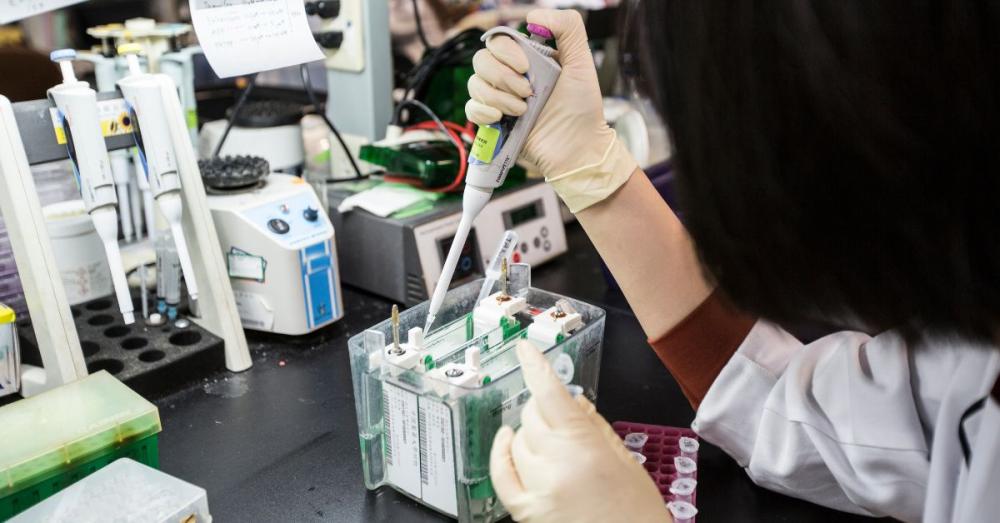
Western blotting, also known as immunoblotting, is a key technique in molecular biology to investigate changes in protein expression in a range of different tissue types. Our detailed Western Blot Protocol walks you through every step, from gel preparation to transfer and detection.
Learn how to:
-
Choose an acrylamide gel percentage, buffer, and blocking solution
-
Select the appropriate loading controls
-
Set up your equipment and consumables
-
Prepare buffers, gel formulations and blocking solutions
-
Carry out electrophoretic transfer and immunoblotting
-
Measure the molecular weight of a protein
-
Quantify protein expression from an immunoblot
Top tip: Don’t underestimate your blocking agent — the wrong one can mask epitopes and ruin your results!
⭐ Find Hello Bio’s full step by step Western Blot Protocol here!
2. Immunocytochemistry (ICC) Protocol
Immunocytochemistry is a type of immunofluorescence technique in which antibodies are used to label cells directly upon either a microscope slide or coverslip. Our ICC Protocol provides a comprehensive guide for fixation, permeabilisation, antibody staining, and imaging.
Learn how to:
-
Choose an isotype control, fixative, and secondary antibody fluorophores
-
Set up your equipment and consumables
-
Prepare solutions and buffers
-
Carry out fixation, antigen retrieval, permeabilisation, and immunofluorescent staining
-
Prepare images for analysis
Top tip: Harsh permeabilisation can destroy epitopes, while too mild a treatment may prevent antibodies from getting in so take particular care with this step!
⭐ Find Hello Bio’s full step by step Immunocytochemistry (ICC) Protocol here!
3. Immunohistochemistry (IHC) Protocol
Immunohistochemistry is an extremely popular and powerful technique that allows the visualisation of protein markers within thin sections of tissue. This can be used to analyse the distribution of receptors, look at the cellular makeup, expression of biomarkers and gross-morphology of a tissue amongst a myriad of other applications. Our IHC Protocol helps you detect protein expression while maintaining tissue architecture.
Learn how to:
-
Choose an isotype control and secondary antibody fluorophores
-
Set up your equipment and consumables for tissue prep, antigen retrieval, free floating and slide mounted detection
-
Prepare tissue for freezing, fixation, and sectioning
-
Prepare solutions and buffers
-
Carry out antigen retrieval and fluorescent & chromogenic detection
Top tip: Tissue fixation can mask epitopes, so optimizing your retrieval method can be the key to success!
⭐ Find Hello Bio’s full step by step Immunohistochemistry (IHC) Protocol here!
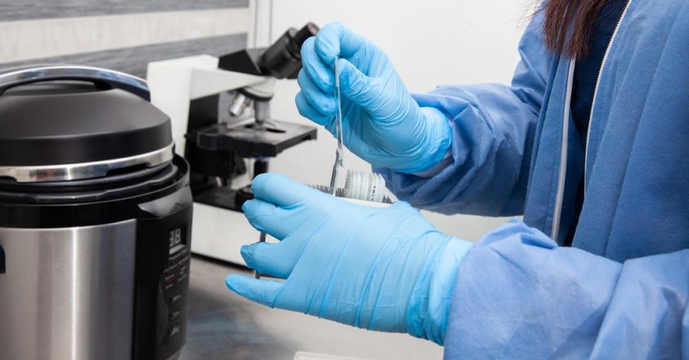
4. Paraffin Embedded Immunohistochemistry Protocol (IHC-P)
Paraffin embedded immunohistochemistry (IHC-P) is a popular variation of IHC where tissue is embedded in paraffin wax following fixation. This better preserves tissue morphology, enables the cutting of extremely thin tissue sections and protects the tissue from degradation (paraffin embedded tissue is stable at room temperature for multiple years). Our IHC-P Protocol covers everything you need to stain sections successfully.
Learn how to:
-
Create buffers, diluents, and solutions
-
Carry out perfusion fixation, immersion fixation, embedding, and sectioning
-
Carry out colorimetric detection using Streptavidin HRP, including deparaffinization, antigen retrieval, blocking, staining, counterstaining, dehydration and mounting
-
Carry out fluorescent detection using deparaffinization, antigen retrieval, blocking, and staining
Top tip: Section thickness can make or break your staining — aim for consistency across your samples.
⭐ Find Hello Bio’s full step by step Paraffin Embedded Immunohistochemistry (IHC-P) Protocol here!
5. Sandwich ELISA Protocol
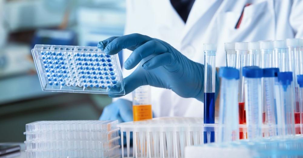
The Enzyme-Linked Immunosorbent Assay (ELISA) is a powerful antibody-based technique for detecting and measuring a specific analyte, such as a protein, within a complex liquid sample. It can be used qualitatively to simply confirm if a target is present, or quantitatively to determine its exact concentration. A key advantage of ELISA over methods like a Western blot or Immunohistochemistry is its ability to provide precise quantitative data for a large number of samples efficiently. It is commonly used to measure biomarkers, hormones, and proteins like cytokines and chemokines. Our Sandwich ELISA Protocol lays out a clear workflow, from plate coating to detection.
Learn how to:
-
Choose between Direct, Indirect, Sandwich and Competitive ELISA
-
Work with colorimetric, fluorescent and chemiluminescence systems
-
Choose an enzyme and substrate pair
-
Set up your equipment and consumables
-
Prepare reagents and buffers
-
Carry out a colorimetric two step detection using a biotinylated detection antibody and streptavidin HRP
-
Analyse the sample data
Top tip: Make sure your standard curve covers the concentration range you expect to measure.
⭐ Find Hello Bio’s full step by step Sandwich ELISA Protocol here!
6. Amyloid-β Protocol
Amyloid Beta (Aβ) is a family of peptides that form the main constituent of senile plaques in Alzheimer's disease and has been extensively investigated for its role in the aetiology of neurodegenerative diseases. If you’re working on Alzheimer’s or neurodegeneration research then our Amyloid-β Protocol is designed to help researchers handle these notoriously tricky peptides.
Learn how to:
-
Handle monomers
-
Monitor the aggregation of Amyloid Beta
-
Preparing oligomers, fibrils and larger insoluble aggregates
-
Separate Aβ aggregates
-
Select the correct use of Amyloid Beta Aggregates
Top tip: Even small changes in peptide concentration or buffer can affect aggregation — consistency is everything.
⭐ Find Hello Bio’s full step by step Amyloid-β Protocol here!
Explore our essential protocol collection
Together, these six protocols cover a wide spectrum of common lab techniques: protein detection, cellular localisation, tissue staining, quantification, and neurodegeneration studies. Whether you’re just starting out or fine-tuning your experiments, they’re designed to save time and give you reliable results.
⭐ Explore the full step-by-step guides at the links above, and don’t forget to bookmark your favourites. Got a protocol you’d like us to feature next? We’d love to hear from you!
Join 1000s of Scientists
Be the first to hear about new product launches, free trials, special offers, and technical tips — straight to your inbox.
Subscribe to the NewsletterHigh Quality – Low Prices
By manufacturing in-house and keeping things simple, we offer savings of up to 50% compared to traditional suppliers — without compromising on quality.


