

Anti-NeuN antibody ValidAb™
Certificate of Analysis
Product overview
| Name | Anti-NeuN antibody ValidAb™ |
| Alternative names | Fox-3 |
| Host | Goat |
| Clonality | Polyclonal |
| Target | NeuN |
| Description | Antibody to NeuN - marker for mature neurones expressed in the nucleus. Part of the ValidAb™ range of highly validated, data-rich antibodies. |
Validation data


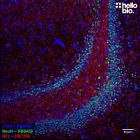
Figure 1. NeuN expressing neurons in the dentate gyrus visualised using HB9493.
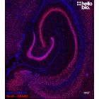
Figure 2. Concentration response of HB9493 staining in rat dentate gyrus.
Figure 3. Independent antibody validation of HB9493 in rat hippocampus.
Product information
| Immunogen | N-terminal 100 amino acids of human FOX3 expressed and purified from E. coli |
| Isotype | IgG |
| Purification | Immunogen affinity purification |
| Concentration | 1mg/ml |
| Formulation | 50% PBS, 50% glycerol + 5mM sodium azide |
| Predicted species reactivity | Mouse, Rat, Human |
| Tested species reactivity | Mouse, Rat |
Tested applications
| Applications | IHC(IF) |
| IHC(IF) optimal concentration | 1:1000 (1μg/ml) as assessed in rat brain sections |
| Positive control | NeuN is highly expressed in the neurons of the CNS and PNS. It is also expressed in SH-SY5Y cells.
|
| Negative control | Any tissue not of neural origin. Most cell lines are NeuN negative. |
| Open data link | Please follow this link to OSF. |
Target information
| Other names | FOX3, RNA binding protein fox-1 homolog 3, Fox-1 homolog C, RBFOX3, RFOX3 |
| UniProt ID | A6NFN3 |
| Structure image |  |
| Gene name | RBFOX3 |
| NCBI full gene name | RNA binding fox-1 homolog 3 |
| Entrez gene ID | 146713 |
| Amino acids | Dependent on isoform |
| Isoforms | NeuN binds primarily to FOX3 which has two isoforms. Isoform 1 is described as the canonical sequence with 312 amino acids (33.8kDa) while isoform 2 has a 13 residue insert at position 312 leading to a total length of 325 amino acids (35.1kDa). NeuN antibodies also bind to synapsin-1 in western blot experiments (but not in IHC or ICC) which has two isoforms. Isoform 1 is 705aa long (74.1kDa) while isoform 2 is shorter at 669aa (70.0kDa). |
| Expression | NeuN is expressed only within neurones. While the vast majority of neurones express NeuN some cell types such as Purkinje cells, stellate and golgi cells do not show immunoreactivity. |
| Subcellular expression | Expression is primarily localised to the nucleus however some FOX3 isoforms can localise to the cytosol. |
| Target function | FOX3 is a splicing regulator of pre-mRNA responsible for neuronal specific alternative splicing of neuronal proteins. |
| Processing | None |
| Post translational modifications | Phosphorylation has been reported (see Lind et al., 2004. J Neurosci Res. 79: 295-302) which is directly related to immunoreactivity whereby dephosphorylation abolished staining. |
| Homology (compared to human) | Mouse FOX3 shows 95.02% identity to human FOX3 wheras rat FOX3 shows no similarity due to a large 47 residue insertion at amino acid 252 in rats. |
| Similar proteins | RNA-binding protein fox-1 homolog 1 (40-44kDa) shows 67.3% identity while RNA-binding protein fox-1 homolog 2 (37-47kDa) shows 56.5% identity. |
Storage & Handling
| Storage instructions | -20°C |
| Shipping Conditions | On ice |
| Important | This product is for RESEARCH USE ONLY and is not intended for therapeutic or diagnostic use. Not for human or veterinary use |
Technical guides
 Western Blot Protocol (1 MB) Immunocytochemistry Protocol (1.2 MB) Immunohistochemistry Protocol (1.6 MB)
 References for Anti-NeuN antibody ValidAb™
-
Novel Insights into NeuN: from Neuronal Marker to Splicing Regulator.
Duan W et al (2016) Molecular neurobiology 53 : 1637-1647 -
NeuN As a Neuronal Nuclear Antigen and Neuron Differentiation Marker.
Gusel'nikova VV et al (2015) Acta naturae 7 : 42-7 -
Identification of neuronal nuclei (NeuN) as Fox-3, a new member of the Fox-1 gene family of splicing factors.
Kim KK et al (2009) The Journal of biological chemistry 284 : 31052-61 -
Characterization of the neuronal marker NeuN as a multiply phosphorylated antigen with discrete subcellular localization.
Lind D et al (2005) Journal of neuroscience research 79 : 295-302 -
NeuN: a useful neuronal marker for diagnostic histopathology.
Wolf HK et al (1996) The journal of histochemistry and cytochemistry : official journal of the Histochemistry Society 44 : 1167-71

Related Products
- Code:
- HB9587
Antibody to MAP2 - cytoskeletal protein used as a neuronal marker. Part of the ValidAb™ range of highly validated, data-rich antibodies.
- Code:
- HB6498
Antibody to NeuN - marker for mature neurones expressed in the nucleus. Part of the ValidAb™ range of highly validated, data-rich antibodies.
- Code:
- HB6429
Antibody to NeuN - marker for mature neurones expressed in the nucleus. Part of the ValidAb™ range of highly validated, data-rich antibodies.