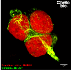
Figure 1. Ki-67 expression in proliferating HEK293T cells
HB7052 reveals the expression of Ki-67 in proliferating HEK293T cells in culture. Method: HEK293T cells were cultured following standard protocols in 10% FBS in DMEM with 1% pen-Strep before being fixed in 4% PFA. Cells were permeabilised with 0.1% Triton X-100 followed by blocking in 1% BSA, 300mM glycine. HB7052 was incubated overnight (4°C) at a 1:1000 dilution (1µg/ml) followed by a one hour incubation with a polyclonal goat anti-mouse DyLight 488 (Thermofisher, 35503) conjugated secondary antibody (1:300 dilution). DAPI (HB0747) was used at 1µg/ml to visualise cell nuclei. For more detail please see our ICC protocol. The image was captured using a Leica SPE confocal laser scanning microscope attached to a Leica DMi8 inverted epifluorescence microscope. The image was captured using a 63x objective and 405nm (24.5% power, PMT: 688V gain) and 488nm (29.0% power, PMT: 775V gain) lasers. Images were captured as a stack (0.23µm z-spacing) before being deconvolved with Huygens professional and flattened using a maximum Z projection in ImageJ (Schindelin et al., 2012. Nat Methods, 9(7), 676–682).
Figure 2. Ki-67 expression is inhibited in serum starved cells but rescued with 20% FBS incubation.
Serum starvation inhibits the ability of HEK293T cells to proliferate therefore inhibits Ki-67 expression. Through stimulation with 20% FBS this then strongly promotes proliferation therefore driving high expression of Ki-67. Method: Following splitting, HEK293T cells were cultured on PLL coated coverslips for 24 hours in 10% FBS in DMEM with 1% Pen/Strep. Cells were washed with plain DMEM and then incubated with DMEM (1% Pen/Strep) for around 20 hours before half of coverslips were then stimulated with 20% FBS for 2hr 45m. Following this all coverslips were then fixed with ice-cold 4% paraformaldehyde in PBS. Cells were permeabilised with 0.1% Triton X-100 followed by blocking in 1% BSA, 300mM glycine. HB7052 was incubated overnight (4°C) at a 1:1000 dilution (1µg/ml) followed by a one hour incubation with a polyclonal goat anti-mouse DyLight 550 (Thermofisher, 84540) conjugated secondary antibody (1:300 dilution) and FITC Phalloidin (HB0814, 1:500 dilution). DAPI (HB0747) was used at 1µg/ml to visualise cell nuclei. For more detail please see our ICC protocol. Images were captured using a Leica DMI6000B inverted epifluorescence microscope attached to a DFC365FX monochrome digital camera. All images were captured using a 40x objective and the same exposure settings of 73.9ms (DAPI), 73ms (FITC Phalloidin) and 107.2ms (Dy550). Images were captured as a stack (0.53µm z-spacing) before being deconvolved in Huygens Professional and flattened using a maximum Z projection in ImageJ (Schindelin et al., 2012. Nat Methods, 9(7), 676–682).
Figure 3. Concentration response of HB7052 staining in HEK293T cells.
HB7502 produces reliable Ki-67 staining at concentrations as low as 1:4,000 (0.25µg/ml). Method: HEK293T cells were cultured following standard protocols in 10% FBS in DMEM with 1% pen-Strep before being fixed in 4% PFA. Cells were permeabilised with 0.1% Triton X-100 followed by blocking in 1% BSA, 300mM glycine. HB7052 was incubated overnight (4°C) at concentrations ranging from 2µg/ml (1:500 dilution) to 0.25µg/ml (1:4,000 dilution). This was followed by a one hour incubation with a polyclonal goat anti-mouse DyLight 488 (Thermofisher, 35503) conjugated secondary antibody (1:300 dilution). DAPI (HB0747) was used at 1µg/ml to visualise cell nuclei. For more detail please see our ICC protocol. Images were captured using a Leica DMI6000B inverted epifluorescence microscope attached to a Photometric-Prime95B monochrome digital camera. Images were captured as a z-stack using a 100x objective with the following exposures:
1:500 – DAPI: 4.9ms, L5: 20ms
1:1,000 – DAPI: 4.9ms, L5: 10ms
1:2,000 – DAPI: 7.9ms, L5: 61ms
1:4,000 – DAPI: 6ms, L5: 15.2ms
Images were deconvolved using Huygens Professional before being flattened using a maximum Z projection in ImageJ (Schindelin et al., 2012. Nat Methods, 9(7), 676–682).
Figure 4. Ki-67 expression in proliferating HEK293T cells
HB7052 reveals the expression of Ki-67 in proliferating HEK293T cells in culture. Method: HEK293T cells were cultured following standard protocols in 10% FBS in DMEM with 1% pen-Strep before being fixed in 4% PFA. Cells were permeabilised with 0.1% Triton X-100 followed by blocking in 1% BSA, 300mM glycine. HB7052 was incubated overnight (4°C) at a 1:1000 dilution (1µg/ml) followed by a one hour incubation with a polyclonal goat anti-mouse DyLight 488 (Thermofisher, 35503) conjugated secondary antibody (1:300 dilution). DAPI (HB0747) was used at 1µg/ml to visualise cell nuclei. For more detail please see our ICC protocol. The image was captured using a Leica SP8 AOBS confocal laser scanning microscope attached to a Leica DMi8 inverted epifluorescence microscope. The image was captured using Lightning adaptive deconvolution using a 63x objective and 405nm (8.6% power, PMT: 800V gain) and 488nm (1.1% power, HyD: 10% gain). Images were captured as a stack (0.38µm z-spacing) before being flattened using a maximum Z projection in ImageJ (Schindelin et al., 2012. Nat Methods, 9(7), 676–682).
Figure 5. Large population of Ki-67 expressing HEK293T cells in culture stained with HB7052.
HB7052 reveals proliferating cells within a large population of HEK293T cells grown in culture. Method: HEK293T cells were cultured following standard protocols in 10% FBS in DMEM with 1% pen-Strep before being fixed in 4% PFA. Cells were permeabilised with 0.1% Triton X-100 followed by blocking in 1% BSA, 300mM glycine. HB7052 was incubated overnight (4°C) at a 1:500 dilution (2µg/ml) followed by a one hour incubation with a polyclonal goat anti-mouse DyLight 488 (Thermofisher, 35503) conjugated secondary antibody (1:300 dilution). DAPI (HB0747) was used at 1µg/ml to visualise cell nuclei. For more detail please see our ICC protocol. The image was captured using a Leica DMI6000B inverted epifluorescence microscope attached to a Photometric-Prime95B monochrome digital camera. The image was captured as a tilescan and z-stack (0.3µm spacing) using a 40x objective, DAPI (4.9ms exposure) and L5 (9.1ms exposure) filters. Images were combined in LASX, deconvolved in Huygens professional then flattened using a maximum Z projection in ImageJ (Schindelin et al., 2012. Nat Methods, 9(7), 676–682).
Figure 6. Ki-67 expression is rescued by 20% FBS stimulation in serum starved HEK293T cells.
Serum starved cells (SS) express only degradation products of Ki-67 whereas 20% FBS stimulation (SS + Stim) induces new Ki-67 expression as seen by the presence of higher molecular weight bands. Method: Following splitting, HEK293T cells were cultured on PLL coated coverslips for 24 hours in 10% FBS in DMEM with 1% Pen/Strep. Cells were washed with plain DMEM and then incubated with DMEM (1% Pen/Strep) for around 20 hours before half of coverslips were then stimulated with 20% FBS for 2hr 45m. Cells were subsequently lysed in Laemmli sample buffer. Rat brain membrane (P2) and cytosol fractions were prepared following previous work (Molnar et al., 1993. Neuroscience 53:307-326) from freshly collected adult brains. Samples were loaded (20µg / lane) onto a 4-20% acrylamide gel alongside a protein ladder (NEB Prestained) before being run at 150V for 50 minutes. Wet transfer to a PVDF membrane was completed in 90 minutes using 400mA. The membrane was blocked for 2hrs in 5% non-fat dry milk before being incubated overnight at 4°C in HB7052 at a 1:500 dilution (2µg/ml). Following washing, the membrane was incubated in secondary antibody (1:10,000 dilution, Polyclonal goat anti-mouse HRP conjugated, Sigma Aldrich A3682) for 2hrs. For more detail please see our Western blotting protocol. Detection was accomplished using Hello Bio ECL and a Licor Odyssey Fc imaging system (ECL channel: 4.5 min exposure, 700nm channel: 30 sec exposure). Following imaging the membrane was stripped with two changes of stripping buffer (HB7756) before being washed, blocked for 2 hours in 5% non-fat dry milk and incubated in HB9177 (mouse monoclonal anti-GAPDH, 1:4,000 dilution, 0.25µg/ml) overnight at 4°C. Following washing, the membrane was incubated in a 1:10,000 dilution of a polyclonal goat anti-mouse HRP conjugated secondary antibody (Sigma Aldrich A3682) for 2hrs and visualized again using Hello Bio ECL and a Licor Odyssey Fc imaging system (ECL channel: 4.5 min exposure, 700nm channel: 30 sec exposure).
Figure 7. Ki-67 expression in the nucleus of a single HEK293T cell revealed with HB7052
HB7052 reveals the subcellular expression of Ki-67 in the nucleus of a proliferating HEK293T cell. Method: HEK293T cells were cultured following standard protocols in 10% FBS in DMEM with 1% pen-Strep before being fixed in 4% PFA. Cells were permeabilised with 0.1% Triton X-100 followed by blocking in 1% BSA, 300mM glycine. HB7052 was incubated overnight (4°C) at a 1:1000 dilution (1µg/ml) followed by a one hour incubation with a polyclonal goat anti-mouse DyLight 488 (Thermofisher, 35503) conjugated secondary antibody (1:300 dilution). DAPI (HB0747) was used at 1µg/ml to visualise cell nuclei. For more detail please see our ICC protocol. The image was captured using a Leica SP8 AOBS confocal laser scanning microscope attached to a Leica DMi8 inverted epifluorescence microscope. The image was captured using Lightning adaptive deconvolution using a 63x objective and 405nm (8.6% power, PMT: 800V gain) and 488nm (3.1% power, HyD: 52.4% gain). Images were captured as a stack (0.12µm z-spacing) before being flattened using a maximum Z projection in ImageJ (Schindelin et al., 2012. Nat Methods, 9(7), 676–682).
Product information
Immunogen
Nuclei derived from the L428 Hodgkin Lymphoma cell line
Clone number
Ki-67
Isotype
IgG1
Purification
Protein A affinity chromatography
Concentration
1mg/ml
Formulation
Lyophilised. When reconstituted contains PBS with 15mM sodium azide and 1% recombinant albumin
Predicted species reactivity
Human, Cow
Tested species reactivity
Human
Tested applications
Applications
ICC, WB
Western blot optimal concentration
2μg/ml (1:500 dilution) as tested in cultured HEK293T cells.
ICC optimal concentration
0.5μg/ml (1:2,000 dilution) as tested in cultured HEK293T cells.
Positive control
All proliferating cells express Ki-67. An easy positive control is using any standard cell line under normal culturing conditions.
Negative control
Cells in G0 do not express Ki-67. An easy negative control is to serum starve cells by growing in serum free media to arrest the cell cycle and inhibit Ki-67 expression.
Ki-67 has two isoforms produced by differential splicing:
Long - 3,256 amino acids (358.7kDa)
Short - 2,896 amino acids (319.4kDa) - missing residues 136-495
Expression
Ki-67 is found in all proliferating cells.
Subcellular expression
Ki-67 is expressed in the nucleus where its localisation changes by cell cycle phase but is predominantly found in association with DNA.
Target function
Ki-67 has multiple important roles in regulating the cell cycle through:
Functionining as a surfactant to stop chromosomes sticking together during mitosis
Mediating attachment of chromosomes to mitotic spindles during mitosis
Involvement in heterochromatin organisation
Processing
Ki-67 is not subject to any processing to form the active conformation.
Post translational modifications
Ki-67 is subject to phosphorylation on many of its serine, threonine and tyrosine residues alongside forming cross-links with SUMO1 and SUMO2 at multiple residues.
Homology (compared to human)
Mouse and rat Ki-67 show low homology with human Ki-67 with homology scores of 42.7% and 43.4% homology respectively. Due to this low homology HB7052 does not react with mouse and rat Ki-67.
Similar proteins
There are no proteins with significant homology to Ki-67
Storage & Handling
Storage instructions
-20°C then use reconstitution advice
Reconstitution advice
Upon receipt store at either -20°C or -80°C.
For 100μg packs either:
Reconstitute with 100μl dH2O and store at 4°C
Reconstitute with 50μl dH2O and 50μl glycerol then store at -20°C
Reconstitute with 100μl dH2O, aliquot then snap freeze and store at -80°C
For 25μg packs either:
Reconstitute with 25μl dH2O and store at 4°C
Reconstitute with 12.5μl dH2O and 12.5μl glycerol then store at -20°C
Reconstitute with 25μl dH2O, aliquot then snap freeze and store at -80°C
For more information read our guide on the best care for your product. Take care when opening as the precipitate is extremely light and can easily be lost if disturbed. When reconstituting make sure that the antibody is thoroughly dissolved by pipetting up and down before giving the antibody a brief spin at 10,000g to make sure that all material is recovered and at the bottom of the tube.
Shipping Conditions
Stable for ambient temperature shipping. Follow storage instructions on receipt.
Important
This product is for RESEARCH USE ONLY and is not intended for therapeutic or diagnostic use. Not for human or veterinary use
Antibody to BrdU - thymidine analogue incorporated into DNA during replication therefore used as a marker of proliferating cells. Part of the ValidAb™ range of highly validated, data-rich antibodies.


 Western Blot Protocol (1 MB)