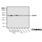
Figure 1. Expression and purification of a GST-fusion protein monitored using HB9897.
A GST-fusion protein was expressed in BL21 E.coli before being purified using glutathione beads with HB9897 being used to check for expression. Method:
A GST-fusion protein was transformed into BL21 E. coli following standard methods (pre-induction)
Protein expression was induced at OD600 = 0.7 with 1mM IPTG (HB3941) and then incubated for 4 hours (post-induction)
Cells were harvested, lysed and the lysate was cleared by spinning at 48,300g for 25 mins (cleared lysate).
The lysate was added to Glutathione Sepharose 4B (Cytiva 17-0756-01) with any unbound being collected (unbound lysate)
The beads were washed to removed undesired proteins (washes 1-4).
The beads were eluted three times with 10mM reduced glutathione (elutions 1-3).
1µl of each sample was loaded onto a 15% acrylamide gel alongside a protein ladder (BioRad Precision Plus Dual Colour, 1610374) before being run at 60V for 40 minutes followed by 120V for 85 minutes. Wet transfer to a PVDF membrane was completed in 90 minutes using 400mA. The membrane was then blocked for 1 hour in 5% non-fat dry milk before being incubated overnight at 4°C in HB9897 (1:5,000 dilution, 0.2µg/ml). Following washing, the membrane was incubated in secondary antibody (1:10,000 dilution, Polyclonal goat anti-mouse HRP conjugated, Sigma Aldrich A3682) for 2hrs. For more detail please see our Western blotting protocol. Detection was accomplished using Clarity Western ECL substrate (BioRad, 1705061) and a Licor Odyssey Fc imaging system (ECL channel: 8 min exposure, 700nm channel: 2 min exposure).
Figure 2. Concentration response of purified GST-tagged protein detected using HB9897.
A GST-fusion protein was expressed in BL21 E.coli before being purified using glutathione beads. HB9897 was able to detect GST-protein expression in as little as 0.01µl of purified lysate. Method: Lysate from a GST-tagged protein preparation was loaded onto a 12% acrylamide gel alongside a protein ladder (BioRad Precision Plus Dual Colour, 1610374) before being run at 60V for 30 minutes followed by 120V for 85 minutes. Wet transfer to a PVDF membrane was completed in 90 minutes using 400mA. The membrane was then blocked for 2 hours in 5% non-fat dry milk before being incubated overnight at 4°C in HB9897 (1:5,000 dilution, 0.2µg/ml). Following washing, the membrane was incubated in secondary antibody (1:10,000 dilution, Polyclonal goat anti-mouse HRP conjugated, Sigma Aldrich A3682) for 2hrs. For more detail please see our Western blotting protocol. Detection was accomplished using Clarity Western ECL substrate (BioRad, 1705061) and a Licor Odyssey Fc imaging system (ECL channel: 10 min exposure, 700nm channel: 2 min exposure).
Figure 3. Concentration response of HB9897 staining in a rat brain cytosol preparation.
HB9897 shows a strong affinity for GST-tagged proteins with protein being visible at as low as a 1 in 64,000 dilution of antibody. Method: A GST-tagged protein was loaded (10µg / lane) onto a 12% acrylamide gel alongside a protein ladder (BioRad Precision Plus Dual Colour, 1610374) before being run at 60V for 30 minutes followed by 120V for 85 minutes. Wet transfer to a PVDF membrane was completed in 90 minutes using 400mA. Following transfer the membrane was cut into strips using Ponceau dye to visualise and cut individual lanes. Strips were blocked for 2hrs in 5% non-fat dry milk before being incubated overnight at 4°C in HB9897. Each strip was incubated separately with a separate HB9897 concentration with this ranging from 1:500 to 1:128,000 dilutions (0.008 - 2µg/ml). Following washing, the membrane was incubated in secondary antibody (1:10,000 Polyclonal goat anti-mouse HRP conjugated, Sigma Aldrich A3682) for 2hrs. For more detail please see our Western blotting protocol. Detection was accomplished using Clarity Western ECL substrate (BioRad, 1705061) and a Licor Odyssey Fc imaging system (ECL channel: 10 min exposure, 700nm channel: 30 sec exposure). Band intensity was calculated using Image Studio version 5.2.5 (LiCor) and a graph was constructed in GraphPad Prism 9 using a 3-parameter Hill equation curve fit.
Figure 4. Detection of as little as 100ng of GST-tagged protein with HB9897.
HB9897 shows a high affinity for GST-tagged proteins with as little as 100ng of protein being visible in a western blot. Method: GST-tagged protein was loaded onto a 12% acrylamide gel alongside a protein ladder (BioRad Precision Plus Dual Colour, 1610374) before being run at 60V for 30 minutes followed by 120V for 90 minutes. Wet transfer to a PVDF membrane was completed in 90 minutes using 400mA. The membrane was then blocked for 2 hours in 5% non-fat dry milk before being incubated overnight at 4°C in HB9897 (1:5,000 dilution, 0.2µg/ml). Following washing, the membrane was incubated in secondary antibody (1:10,000 dilution, Polyclonal goat anti-mouse HRP conjugated, Sigma Aldrich A3682) for 2hrs. For more detail please see our Western blotting protocol. Detection was accomplished using Clarity Western ECL substrate (BioRad, 1705061) and a Licor Odyssey Fc imaging system (ECL channel: 10 min exposure, 700nm channel: 2 min exposure).
Product information
Immunogen
A GST-fusion protein
Clone number
S-tag-05
Isotype
IgG2b
Purification
Protein A affinity chromatography
Concentration
1mg/ml
Formulation
Lyophilised. When reconstituted contains PBS with 15mM sodium azide and 1% recombinant albumin
Predicted species reactivity
Species Independent
Tested species reactivity
Species Independent
Tested applications
Applications
ELISA, WB
Western blot optimal concentration
Dependent upon sample GST-fusion protein expression levels. When 10µg of a GST-fusion protein was loaded a strong signal was observed at antibody concentrations as low as 0.031µg/ml (1:32,000 dilution).
Positive control
Any tissue or cell sample that has been engineered to express a GST-tagged fusion protein.
Glutathione S-transferase class-mu 26 kDa isozyme, Glutathione S-transferase tag
UniProt ID
P08515
Structure image
Amino acids
218 (25.5kDa)
Isoforms
None
Expression
Exogenously expressed only. Not expressed natively in mammalian cells. Expressed natively in the blood fluke Schistosoma japonicum
Subcellular expression
GST-tagged proteins express in a range of subcellular compartments dependent upon the conjugated protein.
Target function
GST-tags serve as an affinity tag that allows for easy purification of a tagged protein using glutathione beads. The GST tag specifically binds to glutathione, facilitating the purification of the target protein by immobilizing it on the beads, while untagged proteins can be washed away, resulting in a highly pure preparation of the GST-tagged protein for downstream analyses.
Storage & Handling
Storage instructions
-20°C then use reconstitution advice
Reconstitution advice
Upon receipt store at either -20°C or -80°C.
For 100μg packs either:
Reconstitute with 100μl dH2O and store at 4°C
Reconstitute with 50μl dH2O and 50μl glycerol then store at -20°C
Reconstitute with 100μl dH2O, aliquot then snap freeze and store at -80°C
For 25μg packs either:
Reconstitute with 25μl dH2O and store at 4°C
Reconstitute with 12.5μl dH2O and 12.5μl glycerol then store at -20°C
Reconstitute with 25μl dH2O, aliquot then snap freeze and store at -80°C
For more information read our guide on the best care for your product. Take care when opening as the precipitate is extremely light and can easily be lost if disturbed. When reconstituting make sure that the antibody is thoroughly dissolved by pipetting up and down before giving the antibody a brief spin at 10,000g to make sure that all material is recovered and at the bottom of the tube.
Shipping Conditions
Stable for ambient temperature shipping. Follow storage instructions on receipt.
Important
This product is for RESEARCH USE ONLY and is not intended for therapeutic or diagnostic use. Not for human or veterinary use
What guarantee do you have that my GST-tag antibody will perform as expected?
We guarantee that your GST-tag antibody will work for the applications and species we list on the datasheet. If the antibody fails to perform as expected then we are happy to offer a 100% refund guarantee. For more details please see our guarantee policy.
What protocols are available for use with this GST-tag antibody?
We have made a comprehensive collection of protocols that we have used in our experiments to validate this GST-tag antibody.
What mounting media do you recommend to use with this antibody?
We recommend using one of our high performance mounting medias, supplied as either hardset or aqeous with a range of counterstains:
What counterstains do you recommend for use in ICC and IHC with this antibody?
We recommend using either DAPI or Hoechst 33342 to label cell nuclei. In some experiments it is also helpful to label actin filaments in the cytoskeleton using a Phalloidin conjugate such as FITC Phalloidin or Rhodamine Phalloidin-TRITC.
Antibody to GFP - green coloured fluorescent protein widely used as a tag in molecular biology. Part of the ValidAb™ range of highly validated, data-rich antibodies.
Monoclonal antibody (IgM) to GFP - green coloured fluorescent protein widely used as a tag in molecular biology. Part of the ValidAb™ range of highly validated, data-rich antibodies.


 Western Blot Protocol (1 MB)