

Super Resolution Microscopy Imaging Tools – high quality, low cost
Janelia Fluor® Preadsorbed Antibodies | Janelia Fluor® Conjugation Kits | Streptavidin Conjugates | Dyes: Amine-Reactive, Click-Active, Free-Acid

Advantages of Janelia Fluor® dyes
- Janelia Fluor® dyes are exceptionally bright, photostable and cell permeable dyes suitable for SRM techniques such as dSTORM and STED as well as live-cell imaging, confocal microscopy, immunocytochemistry and HaloTag® and SNAP-tag® systems.
- Janelia Fluor® dyes are novel fluorescent dyes which are derived from the classic fluorophore tetramethylrhodamine (TMR) with the N,N-dimethyamino groups replaced with azetidine substitutes; a novel, simple modification in the fluorophore structure enabling improved brightness without negatively impacting molecular size and cell permeability.
- Janelia Fluor® conjugates outperform other fluorophores in terms of signal intensity and demonstrate higher resistance to photobleaching.
|
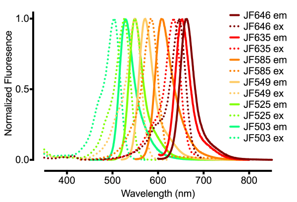
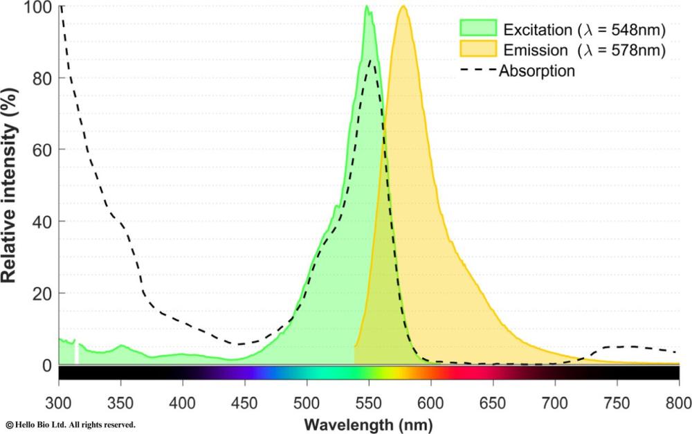
Figure 1. Absorption and emission spectra of Janelia Fluor® dyes. Adapted from Janelia Research Campus [2]. |
Figure 2. Superior anti-fade performance of Janelia Fluor® 549 conjugated dyes compared to those conjugated with DyLight 550 fluorophores. |
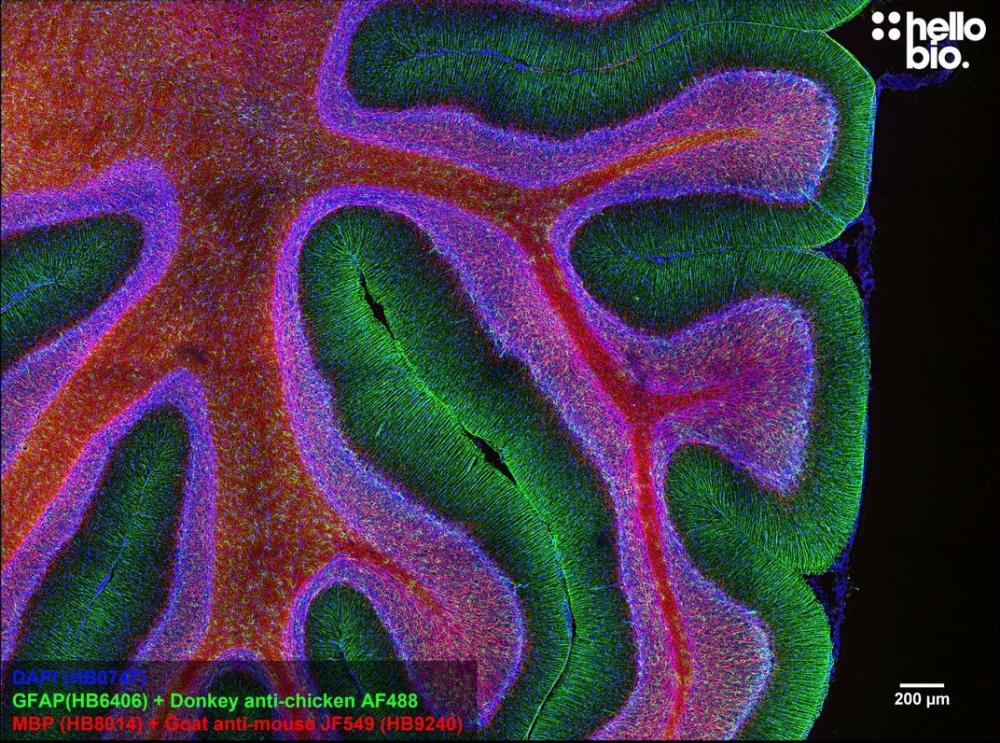
Figure 3. MBP staining in rat cerebellum using Goat Anti-Mouse IgG H&L Janelia Fluor® 549 (HB9240) preadsorbed secondary antibody. |

Super Resolution Imaging Tools Available from Hello Bio
Hello Bio offer a range of high quality yet affordable imaging tools for use in Super Resolution Microscopy (SRM) including:
|
|
||
StreptavidinStreptavidin-based amplification techniques allow for improved detection sensitivity and boosts signal from low-abundance targets. Hello Bio offers a variety of streptavidin conjugates for detection and signal amplification of biotin coupled proteins and antibodies.
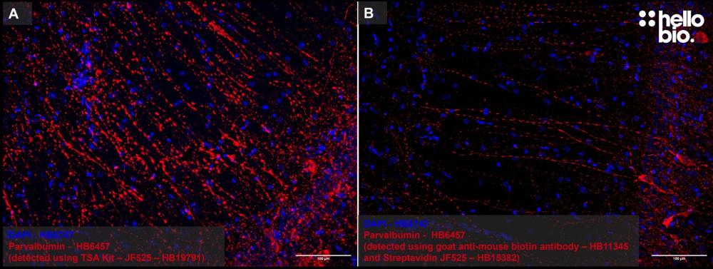
Rat hippocampus stained with parvalbumin anti-mouse biotin antibody followed by incubation with Streptavidin Janelia Fluor® 525 (HB15382) |
|
Biotin Tyramide Signal AmplificationBiotin Tyramide signal amplification (TSA) utilizes horse radish peroxidase (HRP) to enzymatically convert biotin tyramide to bind tyrosine residues on and surrounding the protein epitope targeted by the primary antibody, which can then be detected using streptavidin conjugates. We offer a range of Biotin Tyramide Signal Amplification kits to enhance signal of low abundance targets. Rat hippocampus stained for parvalbumin anti-mouse ValidAbTM antibody detected using Biotin Tyramide Signal Amplification Kit - Janelia Fluor® 525 (HB19791) compared to conjugation with Streptavidin Janelia Fluor® 525 (HB15382). |
DyesHello Bio offer a variety of high quality yet affordable fluorescent dyes to label biomolecules for visualization by fluorescence microscopy, in a range of reactive groups including:
Excitation and emission spectra of Janelia Fluor® 549, free acid (HB8745) |
|
Protocols & Resources:We have a detailed and comprehensive range of protocols and resources to support you: Protocols
Methods & Guides |


Further Reading:
[1] Image adapted from Dr. Eugene Katrukha, Utrecht University, The Netherlands.
[3] Janelia Fluor® dyes: https://www.janelia.org/open-science/janelia-fluor-dyes