

Cresyl Violet Solution (1%)
Certificate of Analysis
Product overview
| Name | Cresyl Violet Solution (1%) |
| Alternative names | Cresyl Echt Violet Solution (0.1%), Cresyl Violet Nissl substance stain solution |
| Biological description | Selectively stains Nissl substance in neurons on formalin fixed, paraffin-embedded tissue. Commonly used for identifying the basic neuronal structure in brain and spinal cord tissue. Nissl granules stain purple/ violet while nuclei of neuroglia and endothelial cells are slightly bluer than Nissl granules (violet to dark blue). Control Tissue: Cerebral Cortex
|
| Description | Stains Nissl substance in neurons on formalin fixed, paraffin-embedded tissue |
Images

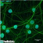
Figure1. Cresyl Violet staining in formalin fixed rat dentate gyrus

Figure2. Cresyl Violet staining in formalin fixed rat prefrontal cortex

Figure3. Cresyl Violet staining in formalin fixed rat CA

Figure4. Cresyl Violet staining in formalin fixed rat hippocampus

Figure5. Cresyl Violet staining in formalin fixed rat cerebellum

Figure6. Cresyl Violet staining in formalin fixed rat cerebellum

Figure7. Cresyl Violet staining in formalin fixed rat prefrontal cortex

Figure8. Cresyl Violet staining in formalin fixed rat cerebellum

Figure9. Cresyl Violet staining in formalin fixed rat cerebellum
Figure1. Cresyl Violet staining in formalin fixed rat dentate gyrus
Figure2. Cresyl Violet staining in formalin fixed rat prefrontal cortex
Figure3. Cresyl Violet staining in formalin fixed rat CA
Figure4. Cresyl Violet staining in formalin fixed rat hippocampus
Figure5. Cresyl Violet staining in formalin fixed rat cerebellum
Figure6. Cresyl Violet staining in formalin fixed rat cerebellum
Figure7. Cresyl Violet staining in formalin fixed rat prefrontal cortex
Figure8. Cresyl Violet staining in formalin fixed rat cerebellum
Figure9. Cresyl Violet staining in formalin fixed rat cerebellum
Biological Data
| Application notes | Protocol: Working Solutions: This stain may be diluted up to 1:10 with deionized water just before use. 1. Deparaffinize and hydrate sections to distilled water. 2. Apply Cresyl Violet Acetate solution (or your Cresyl Violet Acetate working solution) to tissue for 3-5 minutes 3. Quickly rinse in 1 change of distilled water. 4. Dehydrate rapidly in absolute alcohol. Please note that alcohol may remove the stain from tissue over time. 5. Clear in 3 or 4 changes of xylene/xylene substitute and 6. Mount with synthetic resin.
#Protocol 1: Cresyl Violet staining of frozen brain sections
Note: If using paraffin embedded sections, deparaffinise the sections before hydrating into distilled water and then proceed with staining in Cresyl Violet solution |
Solubility & Handling
| Storage instructions | Room temperature |
| Storage of solutions | Prepare and use solutions on the same day if possible. Store solutions at -20°C for up to one month if storage is required. Equilibrate to RT and ensure the solution is precipitate free before use. |
| Shipping Conditions | Stable for ambient temperature shipping. Follow storage instructions on receipt. |
| Important | This product is for RESEARCH USE ONLY and is not intended for therapeutic or diagnostic use. Not for human or veterinary use |
References for Cresyl Violet Solution (1%)
-
Cresyl violet: a superior fluorescent lysosomal marker.
Ostrowski PP et al (2016) Traffic (Copenhagen, Denmark) 17 : 1313-1321 -
Cresyl violet: a red fluorescent Nissl stain.
Alvarez-Buylla A et al (1990) Journal of neuroscience methods 33 : 129-33 -
Cresyl violet: a rapid, simple, easily interpretable stain for detecting Pneumocystis carinii in sputum.
Moas CM et al (1989) Southern medical journal 82 : 957-9


Related Products
- Code:
- HB8199
Blue fluorescent DNA stain. Nuclear counterstain. 1mg/mL staining solution in water. Solid also available in 10mg and 50mg packs.
- Code:
- HB9475