Hello Bio tissue clearing kit enables the easy and quick clearing of tissue and organoids for volumetric imaging without requiring any specialized equipment.
Key features:
Rapid clearing in as little as a day (1mm tissue sections and organoids)
Aqueous (non-solvent) based therefore compatible with standard microscope lenses
Ideal for fluorescent imaging with low quenching after clearing
Contains all key reagents needed including antibody penetration buffer and mounting solution.
Widely compatible with most fluorophores and fluorescent proteins
Preserves cell structures and morphology
No specialized equipment required
Check out our representative 3D reconstructions:
Description
Simple rapid tissue clearing kit for volumetric imaging
Figure 2. Cleared mouse brain segment stained for tyrosine hydroxylase showing dopaminergic projections towards the striatum
Maximum intensity projection of tyrosine hydroxylase staining (HB6605) in a cleared mouse brain segment. Method: A mouse brain was dissected and fixed in 4% PFA for 24 hours before washing and incubation in 30% sucrose for 3 days before being dissected into smaller segments for clearing. The brain segment was incubated in solution A for 3 days at 37°C before washing and incubation in solution B for 3 days. After washing, the tissue was blocked for 3 days then incubated in primary antibody (rabbit polyclonal anti-tyrosine hydroxylase (HB6605)) at a 1:500 dilution for 3 days. Following washing, the tissue was incubated with a goat anti-rabbit DyLight 650 conjugated secondary antibody (1:300 dilution) for a further 3 days. Following more washing the tissue was incubated in mounting solution for 3 days until clear then imaged using a confocal laser scanning microscope to create a z-stack. This was then flattened in ImageJ using the maximum z-projection function. For more information please see our tissue clearing protocol.
Figure 3. Cleared mouse brain lobe stained for tyrosine hydroxylase showing dopaminergic projections towards the striatum
Maximum intensity projection of tyrosine hydroxylase staining (HB6605) in a cleared mouse brain segment. Method: A mouse brain was dissected and fixed in 4% PFA for 24 hours before washing and incubation in 30% sucrose for 3 days before being dissected into smaller segments for clearing. The brain segment was incubated in solution A for 3 days at 37°C before washing and incubation in solution B for 3 days. After washing, the tissue was blocked for 3 days then incubated in primary antibody (rabbit polyclonal anti-tyrosine hydroxylase (HB6605)) at a 1:500 dilution for 3 days. Following washing, the tissue was incubated with a goat anti-rabbit DyLight 650 conjugated secondary antibody (1:300 dilution) for a further 3 days. Following more washing the tissue was incubated in mounting solution for 3 days until clear then imaged using a confocal laser scanning microscope to create a z-stack. This was then flattened in ImageJ using the maximum z-projection function. For more information please see our tissue clearing protocol.
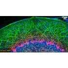
Figure 4. Cleared rat hippocampus stained for GFAP and Neurofilament L
Section of cleared rat hippocampus stained for GFAP (HB8267) and Neurofilament L (HB7266) alongside DAPI counterstaining. Method: Rat hippocampi were dissected from fresh brains and fixed in 4% PFA for 12 hours before washing and incubation in 30% sucrose for 3 days. The hippocampi were incubated in 5ml of solution A for 2 days at 37°C before washing and incubation in 5ml of solution B for 2 days. After washing, the hippocampi were blocked for 3 days then incubated in primary antibodies (mouse anti-GFAP (HB8267) at a 1:200 dilution (5µg/ml) and rabbit anti-neurofilament L (HB7266) at a 1:3,00 dilution (3.3µg/ml) for 1 week. After more washing, the hippocampi were then incubated in secondary antibodies for 6 days (goat anti-mouse Dy488 and goat anti-rabbit Dy594, both at 1:200). Finally, following washing the hippocampi were incubated in DAPI for 3 hours and then washed and incubated in storage solution for 2 days. Images were captured using a Zeiss Z.1 lightsheet microscope. For more information please see our tissue clearing protocol. for 1 week. After more washing, the hippocampi were then incubated in secondary antibodies for 6 days (goat anti-mouse Dy488 and goat anti-rabbit Dy594, both at 1:200). Finally, following washing the hippocampi were incubated in DAPI for 3 hours and then washed and incubated in storage solution for 2 days. Images were captured using a Zeiss Z.1 lightsheet microscope.
Maximum intensity projection of tyrosine hydroxylase staining (HB6605) in a cleared mouse brain segment showing dopaminergic axons in the caudate putamen. Method: A mouse brain was dissected and fixed in 4% PFA for 24 hours before washing and incubation in 30% sucrose for 3 days before being dissected into smaller segments for clearing. The brain segment was incubated in solution A for 3 days at 37°C before washing and incubation in solution B for 3 days. After washing, the tissue was blocked for 3 days then incubated in primary antibody (rabbit polyclonal anti-tyrosine hydroxylase (HB6605)) at a 1:500 dilution for 3 days. Following washing, the tissue was incubated with a goat anti-rabbit DyLight 650 conjugated secondary antibody (1:300 dilution) for a further 3 days. Following more washing the tissue was incubated in mounting solution for 3 days until clear then imaged using a confocal laser scanning microscope to create a z-stack. This was then flattened in ImageJ using the maximum z-projection function. For more information please see our tissue clearing protocol.
Figure 5. Cleared mouse brain segment stained for tyrosine hydroxylase showing dopaminergic neurons in the striatum
Maximum intensity projection of tyrosine hydroxylase staining (HB6605) in a cleared mouse forebrain showing the dopaminergic neurons of the striatum. Method: A mouse brain was dissected and fixed in 4% PFA for 24 hours before washing and incubation in 30% sucrose for 3 days before being dissected into smaller segments for clearing. The brain segment was incubated in solution A for 3 days at 37°C before washing and incubation in solution B for 3 days. After washing, the tissue was blocked for 3 days then incubated in primary antibody (rabbit polyclonal anti-tyrosine hydroxylase (HB6605)) at a 1:500 dilution for 3 days. Following washing, the tissue was incubated with a goat anti-rabbit DyLight 650 conjugated secondary antibody (1:300 dilution) for a further 3 days. Following more washing the tissue was incubated in mounting solution for 3 days until clear then imaged using a confocal laser scanning microscope to create a z-stack. This was then flattened in ImageJ using the maximum z-projection function. For more information please see our tissue clearing protocol.
Figure 1. Representative comparison between HB8771 Tissue Clearing Kit and four other techniques.
Photos were taken before and after mouse brains were cleared following five different tissue clearing protocols:
Figure 2. Cleared mouse brain segment stained for tyrosine hydroxylase showing dopaminergic projections towards the striatum
Maximum intensity projection of tyrosine hydroxylase staining (HB6605) in a cleared mouse brain segment. Method: A mouse brain was dissected and fixed in 4% PFA for 24 hours before washing and incubation in 30% sucrose for 3 days before being dissected into smaller segments for clearing. The brain segment was incubated in solution A for 3 days at 37°C before washing and incubation in solution B for 3 days. After washing, the tissue was blocked for 3 days then incubated in primary antibody (rabbit polyclonal anti-tyrosine hydroxylase (HB6605)) at a 1:500 dilution for 3 days. Following washing, the tissue was incubated with a goat anti-rabbit DyLight 650 conjugated secondary antibody (1:300 dilution) for a further 3 days. Following more washing the tissue was incubated in mounting solution for 3 days until clear then imaged using a confocal laser scanning microscope to create a z-stack. This was then flattened in ImageJ using the maximum z-projection function. For more information please see our tissue clearing protocol.
Figure 3. Cleared mouse brain lobe stained for tyrosine hydroxylase showing dopaminergic projections towards the striatum
Maximum intensity projection of tyrosine hydroxylase staining (HB6605) in a cleared mouse brain segment. Method: A mouse brain was dissected and fixed in 4% PFA for 24 hours before washing and incubation in 30% sucrose for 3 days before being dissected into smaller segments for clearing. The brain segment was incubated in solution A for 3 days at 37°C before washing and incubation in solution B for 3 days. After washing, the tissue was blocked for 3 days then incubated in primary antibody (rabbit polyclonal anti-tyrosine hydroxylase (HB6605)) at a 1:500 dilution for 3 days. Following washing, the tissue was incubated with a goat anti-rabbit DyLight 650 conjugated secondary antibody (1:300 dilution) for a further 3 days. Following more washing the tissue was incubated in mounting solution for 3 days until clear then imaged using a confocal laser scanning microscope to create a z-stack. This was then flattened in ImageJ using the maximum z-projection function. For more information please see our tissue clearing protocol.
Figure 4. Cleared rat hippocampus stained for GFAP and Neurofilament L
Section of cleared rat hippocampus stained for GFAP (HB8267) and Neurofilament L (HB7266) alongside DAPI counterstaining. Method: Rat hippocampi were dissected from fresh brains and fixed in 4% PFA for 12 hours before washing and incubation in 30% sucrose for 3 days. The hippocampi were incubated in 5ml of solution A for 2 days at 37°C before washing and incubation in 5ml of solution B for 2 days. After washing, the hippocampi were blocked for 3 days then incubated in primary antibodies (mouse anti-GFAP (HB8267) at a 1:200 dilution (5µg/ml) and rabbit anti-neurofilament L (HB7266) at a 1:3,00 dilution (3.3µg/ml) for 1 week. After more washing, the hippocampi were then incubated in secondary antibodies for 6 days (goat anti-mouse Dy488 and goat anti-rabbit Dy594, both at 1:200). Finally, following washing the hippocampi were incubated in DAPI for 3 hours and then washed and incubated in storage solution for 2 days. Images were captured using a Zeiss Z.1 lightsheet microscope. For more information please see our tissue clearing protocol. for 1 week. After more washing, the hippocampi were then incubated in secondary antibodies for 6 days (goat anti-mouse Dy488 and goat anti-rabbit Dy594, both at 1:200). Finally, following washing the hippocampi were incubated in DAPI for 3 hours and then washed and incubated in storage solution for 2 days. Images were captured using a Zeiss Z.1 lightsheet microscope.
Maximum intensity projection of tyrosine hydroxylase staining (HB6605) in a cleared mouse brain segment showing dopaminergic axons in the caudate putamen. Method: A mouse brain was dissected and fixed in 4% PFA for 24 hours before washing and incubation in 30% sucrose for 3 days before being dissected into smaller segments for clearing. The brain segment was incubated in solution A for 3 days at 37°C before washing and incubation in solution B for 3 days. After washing, the tissue was blocked for 3 days then incubated in primary antibody (rabbit polyclonal anti-tyrosine hydroxylase (HB6605)) at a 1:500 dilution for 3 days. Following washing, the tissue was incubated with a goat anti-rabbit DyLight 650 conjugated secondary antibody (1:300 dilution) for a further 3 days. Following more washing the tissue was incubated in mounting solution for 3 days until clear then imaged using a confocal laser scanning microscope to create a z-stack. This was then flattened in ImageJ using the maximum z-projection function. For more information please see our tissue clearing protocol.
Figure 5. Cleared mouse brain segment stained for tyrosine hydroxylase showing dopaminergic neurons in the striatum
Maximum intensity projection of tyrosine hydroxylase staining (HB6605) in a cleared mouse forebrain showing the dopaminergic neurons of the striatum. Method: A mouse brain was dissected and fixed in 4% PFA for 24 hours before washing and incubation in 30% sucrose for 3 days before being dissected into smaller segments for clearing. The brain segment was incubated in solution A for 3 days at 37°C before washing and incubation in solution B for 3 days. After washing, the tissue was blocked for 3 days then incubated in primary antibody (rabbit polyclonal anti-tyrosine hydroxylase (HB6605)) at a 1:500 dilution for 3 days. Following washing, the tissue was incubated with a goat anti-rabbit DyLight 650 conjugated secondary antibody (1:300 dilution) for a further 3 days. Following more washing the tissue was incubated in mounting solution for 3 days until clear then imaged using a confocal laser scanning microscope to create a z-stack. This was then flattened in ImageJ using the maximum z-projection function. For more information please see our tissue clearing protocol.
Prepare and use solutions on the same day if possible. Store solutions at -20°C for up to one month if storage is required. Equilibrate to RT and ensure the solution is precipitate free before use.
Handling
Please note that some of the solutions require warming to 37°C and any precipitate redissolving before use
Shipping Conditions
Stable for ambient temperature shipping. Follow storage instructions on receipt.
Important
This product is for RESEARCH USE ONLY and is not intended for therapeutic or diagnostic use. Not for human or veterinary use.
Is this tissue clearing kit solvent or aqueous based
The kit is aqeuous based therefore is non-toxic and compatible with imaging systems.
My solution(s) have precipitates in them, is this normal?
Many solutions contain high solute concentrations which tend to precipitate at lower temperatures. We recommend incubating at 37°C for 1 hour and stirring before use.
What is the refractive index of the mounting and storage solution?
The mounting and storage solution has a refractive index of 1.46


 Tissue clearing protocol book (316.6 KB)