

Anti-GFAP antibody ValidAb™
Certificate of Analysis
Product overview
| Name | Anti-GFAP antibody ValidAb™ |
| Host | Goat |
| Clonality | Polyclonal |
| Target | GFAP |
| Description | Antibody to GFAP - cytoskeletal protein used as an astrocyte marker. Part of the ValidAb™ range of highly validated, data-rich antibodies. |
Validation data


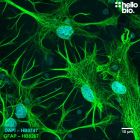
Figure 1. Astrocytes in culture in a cultured rat neuron preparation.
Figure 2. Astrocyte populations in culture stained by HB7592.
Figure 3. Astrocytes stained for GFAP with HB7592 in the cerebellum
Figure 4. Independent antibody validation of HB7592 and HB6406.
Figure 5. Concentration response of HB7592 staining in cultured rat neurons.
Product information
| Immunogen | Recombinant human GFAP (isoform 1) expressed in and purified from E. coli |
| Purification | Immunogen affinity purification |
| Concentration | 1mg/ml |
| Formulation | 50% PBS, 50% glycerol + 5mM sodium azide |
| Predicted species reactivity | Mouse, Rat, Human |
| Tested species reactivity | Mouse, Rat |
Tested applications
| Applications | ICC, IHC(IF) |
| IHC(IF) optimal concentration | 0.5µg/ml (1:2,000 dilution) as tested in free-floating paraformaldehyde fixed rat brain sections |
| ICC optimal concentration | 0.5µg/ml (1:2,000 dilution) as tested in cultured rat neurones |
| Positive control | GFAP is highly expressed in neural tissues containing astrocytes. It is not widely expressed in cell lines, however it is in specific lines such as U-87 MG. |
| Negative control | Most non-neural tissues. |
| Open data link | Please follow this link to OSF |
Target information
| UniProt ID | P14136 |
| Structure image |  |
| Gene name | GFAP |
| NCBI full gene name | glial fibrillary acidic protein |
| Entrez gene ID |
| Amino acids | 432 (49.9kDa) |
| Isoforms | GFAP has three confirmed and 21 potential isoforms. Isoform 1 (GFAP alpha): canonical, 49.9kDa; Isoform 2 (GFAP epsilon): amino acid changes between positions 391 and 432, 49.5kDa; Isoform 3 (GFAP kappa): amino acid changes between positions 391 and 432, 50.3kDa |
| Expression | GFAP is primarily expressed within astrocytes of the central nervous system alongside also expressing in non-myelinating Schwann cells of the peripheral nervous system and satellite cells of the peripheral ganglia. GFAP expression has also been reported in Leydig cells of the testis alongside stellate cells from the pancreas and liver in rats. |
| Subcellular expression | GFAP is a key cytoskeletal component therefore is widely expressed as bundles of GFAP positive fibres. |
| Target function | GFAP is a class III intermediate filament protein with an important role in many processes beyond being a structural cytoskeletal component. GFAP is involved in mitosis of astrocytres, mediating interactions between astrocytes and neurones and repair after CNS injury. |
| Processing | Following translation, no processing is required for GFAP to reach its active conformation. |
| Post translational modifications | GFAP is subjected to numerous post-translational modifications including 9 phosphorylation sites which are the target of AURKB and ROCK1 alongside 5 separate citrullination sites. |
| Homology (compared to human) | Rat, mouse and human GFAP proteins have a 90% similarity score in a direct BLAST comparison. |
| Similar proteins | Other type III intermediate filament proteins have homology with GFAP including Vimentin (58%), Desmin (59%) and Peripherin (56%) when assessed using BLAST. |
Storage & Handling
| Storage instructions | -20°C |
| Shipping Conditions | On ice |
| Important | This product is for RESEARCH USE ONLY and is not intended for therapeutic or diagnostic use. Not for human or veterinary use |
Technical guides
 Western Blot Protocol (1 MB) Immunocytochemistry Protocol (1.2 MB) Immunohistochemistry Protocol (1.6 MB)
 References for Anti-GFAP antibody ValidAb™
-
Importance of GFAP isoform-specific analyses in astrocytoma.
van Bodegraven EJ et al (2019) Glia 67 : 1417-1433 -
The role of GFAP and vimentin in learning and memory.
Wilhelmsson U et al (2019) Biological chemistry 400 : 1147-1156 -
GFAP-expressing progenitors are the principal source of constitutive neurogenesis in adult mouse forebrain.
Garcia AD et al (2004) Nature neuroscience 7 : 1233-41 -
Glial fibrillary acidic protein: GFAP-thirty-one years (1969-2000).
Eng LF et al (2000) Neurochemical research 25 : 1439-51 -
GFAP and astrogliosis.
Eng LF et al (1994) Brain pathology (Zurich, Switzerland) 4 : 229-37


Related Products
- Code:
- HB8001
Antibody to GFAP - cytoskeletal protein used as an astrocyte marker. Part of the ValidAb™ range of highly validated, data-rich antibodies.
- Code:
- HB8267
Antibody to GFAP - cytoskeletal protein used as an astrocyte marker. Part of the ValidAb™ range of highly validated, data-rich antibodies.
- Code:
- HB6406
Antibody to GFAP - cytoskeletal protein used as an astrocyte marker. Part of the ValidAb™ range of highly validated, data-rich antibodies.