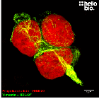
D-luciferin sodium salt is a water-soluble chemiluminescent luciferase substrate which is commonly used for in vivo imaging of the expression of luciferase. It is also frequently used in vitro.
The compound is nontoxic and stable in cells and live animals.
The luciferin substrate can be oxidized by the luciferase enzyme to generate an excited state molecule that emits light.
D-Luciferin sodium salt product vial image | Hello Bio
D-Luciferin sodium salt product vial image | Hello Bio
Solubility & Handling
Storage instructions
-20°C
Solubility overview
Soluble in water (100 mM), and in DMSO (100 mM)
Storage of solutions
Prepare and use solutions on the same day if possible. Store solutions at -20°C for up to one month if storage is required. Equilibrate to RT and ensure the solution is precipitate free before use.
Shipping Conditions
Stable for ambient temperature shipping. Follow storage instructions on receipt.
Important
This product is for RESEARCH USE ONLY and is not intended for therapeutic or diagnostic use. Not for human or veterinary use
Dynamic bioluminescence imaging for quantitative tumour burden assessment using IV or IP administration of D: -luciferin: effect on intensity, time kinetics and repeatability of photon emission.
Keyaerts et al (2008) Eur J Nucl Med Mol Imaging. 35(5) : 999-1007


![D-Luciferin sodium salt | [103404-75-7] Chemical Structure](https://cdn.hellobio.com/media/catalog/product//h/b/hb5119_1.png "D-Luciferin sodium salt | [103404-75-7]")
 Understanding purity and quality - a guide for life scientists