Antibody to Histone H3 - Histone protein used as a loading control for western blotting. Part of the ValidAb™ range of highly validated, data-rich antibodies.
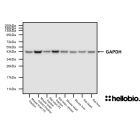
Figure 1. Histone H3 expression in various tissue lysates and preparations.
HB6980 recognises the 14kDa and 16kDa isoforms of Histone H3 in a range of tissues and lysates. Method: mouse brain and rat brain membrane (P2) and cytosol fractions were prepared following previous work (Molnar et al., 1993. Neuroscience 53:307-326) from freshly collected adult brains. Other tissue lysates were prepared following established protocols from freshly dissected tissue (see our guide on WB sample preparation). Samples were loaded (20µg / lane) onto a 20% acrylamide gel alongside a protein ladder (BioRad Precision Plus Dual Colour) before being run at 60V for 30 minutes followed by 100V for 80 minutes then 130V for 40 minutes. Wet transfer to a PVDF membrane was completed in 90 minutes using 400mA. The membrane was blocked for 2hrs in 5% non-fat dry milk before being incubated overnight at 4°C in HB6980 at a 1:1,000 dilution. Following washing, the membrane was incubated in secondary antibody (1:10,000 dilution, Polyclonal goat anti-rabbit HRP conjugated, Sigma, A6154) for 2hrs. For more detail please see our Western blotting protocol. Detection was accomplished using Clarity Western ECL substrate (BioRad, 1705061) and a Licor Odyssey Fc imaging system (ECL channel: 6 min exposure, 700nm channel: 30 sec exposure). Following imaging the membrane was stripped with two changes of stripping buffer (HB7756) before being washed, blocked for 2 hours in 5% non-fat dry milk and incubated in HB9177 (mouse monoclonal anti-GAPDH, 1:4,000 dilution, 0.25µg/ml) and HB8224 (rabbit polyclonal anti-ß-actin, 1:1,000 dilution) overnight at 4°C. Following washing the membrane was incubated in a 1:10,000 dilution of polyclonal goat anti-mouse HRP conjugated (Sigma Aldrich A3682) and polyclonal goat anti-rabbit HRP (Sigma Aldrich A6154) conjugated secondary antibodies for 2hrs and visualised again using Clarity Western ECL substrate (BioRad, 1705061) and a Licor Odyssey Fc imaging system (ECL channel: 4 min exposure, 700nm channel: 30 sec exposure).
Figure 2. Concentration response of HB6980 staining in a rat brain cytosol preparation.
HB6980 shows strong affinity for Histone H3 with bands visible at as low a concentration as 8.75ng/ml (1:64,000 dilution). Method: cytosol fractions were prepared from fresh rat brains following established protocols (Molnar et al., 1993. Neuroscience 53:307-326). Rat cytosol samples were loaded (20µg / lane) onto a 20% acrylamide gel alongside a protein ladder (BioRad Precision Plus Dual Colour) before being run at 60V for 30 minutes followed by 120V for 100 minutes. Wet transfer to a PVDF membrane was completed in 90 minutes using 400mA. Following transfer the membrane was cut into strips using Ponceau dye to visualise and cut individual lanes. Strips were blocked for 2hrs in 5% non-fat dry milk before being incubated overnight at 4°C in HB6980. Each strip was incubated separately with a separate HB6980 concentration with this ranging from 1:500 to 1:128,000 dilutions (4.38 – 1120ng/ml). Following washing, the membrane was incubated in secondary antibody (1:10,000 dilution, Polyclonal goat anti-rabbit HRP conjugated, Sigma, A6154) for 2hrs. For more detail please see our Western blotting protocol. Detection was accomplished using Clarity Western ECL substrate (BioRad, 1705061) and a Licor Odyssey Fc imaging system (ECL channel: 8 min exposure, 700nm channel: 30 sec exposure). Band intensity was calculated using Image Studio version 5.2.5 (LiCor) and a graph was constructed in GraphPad Prism 9 using a 3-parameter Hill equation curve fit.
Product information
Immunogen
Synthetic peptide consisting of residues from within the C-terminus of human Histone H3. The sequence of the immunogen is: REIRRYQKSTELLIRKLPFQRLMREIAQDFKTDLRFQSSAVMALQEACESYLVGLFEDTNLCVIHAKRVTIMPKDIQLARRIRGERA
Isotype
IgG
Purification
Immunogen affinity chromatography
Concentration
0.56 mg/mL
Formulation
PBS with 0.01% thiomersal and 50% glycerol, pH7.3
Predicted species reactivity
Mouse, Rat, Human
Tested species reactivity
Mouse, Rat
Tested applications
Applications
WB
Western blot optimal concentration
280ng/ml (1:2,000 dilution) as tested in a rat brain cytosol preparation
Positive control
Histone H3 is found in the nucleus of all cell types therefore any tissue or cell line can serve as a positive control.
Negative control
Any negative control tissues or cells must not contain any nuclei therefore red blood cells or platelets can work well in adittion to samples that have undergone subcellular fractionation to remove any nuclei.
Histone H3 has 21 family members with this antibody having an immunogen based upon Histone H3.1t. This family member has no known isoforms. Please note that this antibody is not isoform specific.
Expression
Histone H3 is a ubiquitous protein that is expressed in almost all eukaryotic cells throughout the body. It is found in the nuclei of cells in various tissues and organs, including but not limited to the brain, heart, liver, lungs, kidneys, and skin.
Subcellular expression
Histone H3 proteins are expressed in the nucleus where they form the structure of chromatin around which DNA wounds.
Target function
Histone H3 is a key protein involved in the organization and packaging of DNA into a compact structure called chromatin in eukaryotic cells. It plays a critical role in regulating gene expression by determining the accessibility of DNA to transcription factors and other regulatory proteins. Additionally, histone H3 is subject to various post-translational modifications, such as methylation, acetylation, and phosphorylation, which are important for epigenetic regulation and influence chromatin structure and function.
Processing
Prior to forming an active conformation the initiator methionine is removed.
Post translational modifications
Histone H3 proteins are subject to numerous post-translational modifications which mediate epigenetic control of gene expression. These modifications include methylation, acetylation and phosphorylation on multiple residues amongst other modifications.
Homology (compared to human)
Compared to human Histone H3.1t:
Histone H3.1 was the closest homologue in mice with a 97.1% identity score
Histone H3.1t was the closest homologue in rats with a 97.1% identity score.
Similar proteins
In a BLAST search no non-Histone H3 family members were identified as having significant homology with Histone H3.1t
Epitope homology (between species)
A BLAST search of the immunogen reveals a 96.55% homology with mouse and rat Histone H3.1
Epitope homology (other proteins)
A BLAST search of the immunogen shows that the only proteins with homology to it are Histone H3 family members.
Storage & Handling
Storage instructions
-20°C
Shipping Conditions
On ice
Important
This product is for RESEARCH USE ONLY and is not intended for therapeutic or diagnostic use. Not for human or veterinary use
Antibody to Histone H3 - Histone protein used as a loading control for western blotting. Part of the ValidAb™ range of highly validated, data-rich antibodies.
Antibody to β-tubulin - cytoskeletal component widely used for imaging microtubules and as a loading control. Part of the ValidAb™ range of highly validated, data-rich antibodies.


 Western Blot Protocol (1 MB)