

aCSF Instant Powder (packets)
Certificate of Analysis
Product overview
| Name | aCSF Instant Powder (packets) |
| Biological description | Artificial cerebrospinal fluid (aCSF) is a widely used buffer in electrophysiological experiments to sustain ex-vivo brain sections. This kit contains 20 instant powder packets. Simply dissolve the contents of each packet in dH2O to a final volume of 1L, mix and bubble with carbogen to make 1L of aCSF at physiological pH.
Key features:
Contains (in mM): NaCl 124. Glucose 10, NaHCO3 24, KCl 3, NaH2PO4 1.25, CaCl2 2.5, MgCl2 1.3
|
| Customer comments | Amazing. We were having issues getting consistency with our 2-photon slice imaging and ultimately it must have been the osmolarity of our previous aCSF that we were making because the minute we switched to Hello Bio, our results just got better. Honestly, so so worth it!! And really easy and straightforward. I love this product. Verified customer, MIT |
| Description | Preformulated instant powder packets to make artificial cerebrospinal fluid (aCSF) |
Images

Figure 1. Representative traces of different experiment types carried out in HB9200 aCSF
Plateau potentials: Cells were held using current clamp and adjusted to a resting voltage of -69mV. Cells were then stimulated with one pulse followed by 5 stimuli at 100Hz frequency.
EPSP: Cells were held using current clamp and adjusted to a resting voltage of -69mV before being stimulated with a single pulse.
IPSC: Cells were held at 0mV using voltage clamp before being stimulated with a single pulse.
EPSC: Cells were held at -70mV before being stimulated via a paired pulse protocol with an interval of 50ms.

Figure 2. Spontaneous EPSC and IPSC currents recorded using HB9200 aCSF

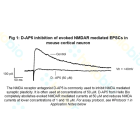
Figure 3. Long term recording of EPSCs in HB9200 aCSF for over 70 minutes
 (HB9200) manufactured by Hello Bio")
Figure 4. aCSF Instant Powder (packets) (HB9200) manufactured by Hello Bio
Figure 1. Representative traces of different experiment types carried out in HB9200 aCSF
Plateau potentials: Cells were held using current clamp and adjusted to a resting voltage of -69mV. Cells were then stimulated with one pulse followed by 5 stimuli at 100Hz frequency.
EPSP: Cells were held using current clamp and adjusted to a resting voltage of -69mV before being stimulated with a single pulse.
IPSC: Cells were held at 0mV using voltage clamp before being stimulated with a single pulse.
EPSC: Cells were held at -70mV before being stimulated via a paired pulse protocol with an interval of 50ms.
Figure 2. Spontaneous EPSC and IPSC currents recorded using HB9200 aCSF
Figure 3. Long term recording of EPSCs in HB9200 aCSF for over 70 minutes
Figure 4. aCSF Instant Powder (packets) (HB9200) manufactured by Hello Bio
Solubility & Handling
| Storage instructions | RT. Add each packet to 1L dH2O. |
| Storage of solutions | Prepare and use solutions on the same day if possible. Store solutions at -20°C for up to one month if storage is required. Equilibrate to RT and ensure the solution is precipitate free before use. |
| Handling | Dissolve the contents of each packet in dH2O to a final volume of 1000ml, mix well and bubble with carbogen (10-15 minutes) to make 1L of aCSF at physiological pH. Warm to 37°C before use. Use immediately once opened. |
| Shipping Conditions | Stable for ambient temperature shipping. Follow storage instructions on receipt. |
| Important | This product is for RESEARCH USE ONLY and is not intended for therapeutic or diagnostic use. Not for human or veterinary use |
Chemical Data
| Kit contents | Preformulated packets. Each makes 1L of aCSF. |
| pH after carbogenation | 7.2 |
| pH before carbogenation | 7.5 |
References for aCSF Instant Powder (packets)
-
Reduced expression of the psychiatric risk gene DLG2 (PSD93) impairs hippocampal synaptic integration and plasticity.
Griesius S et al (2022) Neuropsychopharmacology : official publication of the American College of Neuropsychopharmacology 47 : 1367-1378 -
The development of synaptic plasticity induction rules and the requirement for postsynaptic spikes in rat hippocampal CA1 pyramidal neurones.
Buchanan KA et al (2007) The Journal of physiology 585 : 429-45


New Products in this Area
Preformulated instant powder packets to make artificial cerebrospinal fluid (aCSF) without Mg2+ or Ca2+
Preformulated packets to make cutting solution for electrophysiology.
Preformulated instant powder packets to make artificial cerebrospinal fluid (aCSF) without Mg2+ or Ca2+
Related Products
- Code:
- HB0225
Selective, competitive NMDA receptor antagonist. Inhibits NMDAR-synaptic plasticity.
- Code:
- HB0443
- Code:
- HB4822