

aCSF Instant Powder (Mg2+/Ca2+ free) (packets)
Certificate of Analysis
Product overview
| Name | aCSF Instant Powder (Mg2+/Ca2+ free) (packets) |
| Biological description | Artificial cerebrospinal fluid (aCSF) is a widely used buffer in electrophysiological experiments to sustain ex-vivo brain sections. This kit contains 20 instant powder packets. Simply dissolve the contents of each packet in dH2O to a final volume of 1L, mix, add the desired concentration of Mg2+ and Ca2+ and bubble with carbogen to make 1L of aCSF at physiological pH. Please note: This formulation does not contain any Mg2+ or Ca2+ so that this can be specified by the experimenter.
Key features:
Contains (in mM): NaCl 124. Glucose 10, NaHCO3 24, KCl 3, NaH2PO4 1.25 |
| Description | Preformulated instant powder packets to make artificial cerebrospinal fluid (aCSF) without Mg2+ or Ca2+ |
Images
")
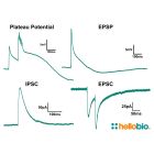
Figure 1. Representative traces of different experiment types carried out in aCSF Instant Powder (packets)
Representative traces from four different experiments to measure NMDA dependent plateau potentials, excitatory post synaptic potentials (EPSPs), inhibitory postsynaptic currents (IPSCs) and excitatory postsynaptic currents (EPSCs). Method: C57BL/6J mouse brain sections were prepared following standard protocols (Udakis et al., 2020). Pyramidal neurons in hippocampal CA1 were patched using a CsMeSO4 internal solution containing QX314 and stimulated via the stratum radiatum to excite Schaffer collateral synapses.
Plateau potentials: Cells were held using current clamp and adjusted to a resting voltage of -69mV. Cells were then stimulated with one pulse followed by 5 stimuli at 100Hz frequency.
EPSP: Cells were held using current clamp and adjusted to a resting voltage of -69mV before being stimulated with a single pulse.
IPSC: Cells were held at 0mV using voltage clamp before being stimulated with a single pulse.
EPSC: Cells were held at -70mV before being stimulated via a paired pulse protocol with an interval of 50ms.
")
Figure 2. Spontaneous EPSC and IPSC currents recorded using aCSF Instant Powder (packets)
Traces showing both spontaneous excitatory postsynaptic currents (EPSCs) and spontaneous inhibitory postsynaptic currents (IPSCs). Method: C57BL/6J mouse brain sections were prepared following standard protocols (Udakis et al., 2020). Pyramidal neurons in hippocampal CA1 were patched using a CsMeSO4 internal solution containing QX314. Neurons were held in voltage clamp with sEPSCs being recorded at -70mV and sIPSCs being recorded at 0mV.
 for over 70 minutes")
Figure 3. Long term recording of EPSCs in aCSF Instant Powder (packets) for over 70 minutes
Summary of experiment recording excitatory post synaptic currents (EPSCs) for over 75 minutes in mouse pyramidal neurons. Method: C57BL/6J mouse brain sections were prepared following standard protocols (Udakis et al., 2020). Pyramidal neurons in hippocampal CA1 were patched using a CsMeSO4 internal solution containing QX314, held at -70mV in voltage clamp and stimulated in a paired pulse stimulation protocol (50ms interval) via the stratum radiatum to excite Schaffer collateral synapses. Figures shown are representative traces from different timepoints during the experiment, the input current required to hold the cell at -70mV, the series resistance, the amplitude of the initial EPSC and the ratio of amplitudes of the two EPSCs (paired pulse ratio).
Figure 1. Representative traces of different experiment types carried out in aCSF Instant Powder (packets)
Representative traces from four different experiments to measure NMDA dependent plateau potentials, excitatory post synaptic potentials (EPSPs), inhibitory postsynaptic currents (IPSCs) and excitatory postsynaptic currents (EPSCs). Method: C57BL/6J mouse brain sections were prepared following standard protocols (Udakis et al., 2020). Pyramidal neurons in hippocampal CA1 were patched using a CsMeSO4 internal solution containing QX314 and stimulated via the stratum radiatum to excite Schaffer collateral synapses.
Plateau potentials: Cells were held using current clamp and adjusted to a resting voltage of -69mV. Cells were then stimulated with one pulse followed by 5 stimuli at 100Hz frequency.
EPSP: Cells were held using current clamp and adjusted to a resting voltage of -69mV before being stimulated with a single pulse.
IPSC: Cells were held at 0mV using voltage clamp before being stimulated with a single pulse.
EPSC: Cells were held at -70mV before being stimulated via a paired pulse protocol with an interval of 50ms.
Figure 2. Spontaneous EPSC and IPSC currents recorded using aCSF Instant Powder (packets)
Traces showing both spontaneous excitatory postsynaptic currents (EPSCs) and spontaneous inhibitory postsynaptic currents (IPSCs). Method: C57BL/6J mouse brain sections were prepared following standard protocols (Udakis et al., 2020). Pyramidal neurons in hippocampal CA1 were patched using a CsMeSO4 internal solution containing QX314. Neurons were held in voltage clamp with sEPSCs being recorded at -70mV and sIPSCs being recorded at 0mV.
Figure 3. Long term recording of EPSCs in aCSF Instant Powder (packets) for over 70 minutes
Summary of experiment recording excitatory post synaptic currents (EPSCs) for over 75 minutes in mouse pyramidal neurons. Method: C57BL/6J mouse brain sections were prepared following standard protocols (Udakis et al., 2020). Pyramidal neurons in hippocampal CA1 were patched using a CsMeSO4 internal solution containing QX314, held at -70mV in voltage clamp and stimulated in a paired pulse stimulation protocol (50ms interval) via the stratum radiatum to excite Schaffer collateral synapses. Figures shown are representative traces from different timepoints during the experiment, the input current required to hold the cell at -70mV, the series resistance, the amplitude of the initial EPSC and the ratio of amplitudes of the two EPSCs (paired pulse ratio).
Solubility & Handling
| Storage instructions | RT. Dissolve each pack in dH2O to 1L final volume. |
| Storage of solutions | Prepare and use solutions on the same day if possible. Store solutions at -20°C for up to one month if storage is required. Equilibrate to RT and ensure the solution is precipitate free before use. |
| Handling | Dissolve the contents of each packet in dH2O, add the desired quantity of Mg2+ and Ca2+ then bring the final volume to 1000ml and bubble with carbogen (10-15 minutes) to make 1L of aCSF at physiological pH. Warm to 37°C before use. Use immediately once opened. |
| Shipping Conditions | Stable for ambient temperature shipping. Follow storage instructions on receipt. |
| Important | This product is for RESEARCH USE ONLY and is not intended for therapeutic or diagnostic use. Not for human or veterinary use |
Chemical Data
| Kit contents | Preformulated packets. Each makes 1L of aCSF. |
| pH after carbogenation | 7.2 |
| pH before carbogenation | 7.5 |
References for aCSF Instant Powder (Mg2+/Ca2+ free) (packets)
-
Reduced expression of the psychiatric risk gene DLG2 (PSD93) impairs hippocampal synaptic integration and plasticity.
Griesius S et al (2022) Neuropsychopharmacology : official publication of the American College of Neuropsychopharmacology 47 : 1367-1378 -
The development of synaptic plasticity induction rules and the requirement for postsynaptic spikes in rat hippocampal CA1 pyramidal neurones.
Buchanan KA et al (2007) The Journal of physiology 585 : 429-45

New Products in this Area
Preformulated packets to make cutting solution for electrophysiology.
Related Products
- Code:
- HB9200
Preformulated instant powder packets to make artificial cerebrospinal fluid (aCSF)
- Code:
- HB0225
Selective, competitive NMDA receptor antagonist. Inhibits NMDAR-synaptic plasticity.