SuperBlotTM ECL Western Blotting Substrate Kit (Standard)

(HB7090)
Technical documents: Datasheet

Product overview

Name SuperBlotTM ECL Western Blotting Substrate Kit (Standard)
Biological description

Overview

Hello Bio SuperBlotTM ECL Western Blotting Substrate Kit (Standard) is an enhanced chemiluminescent (ECL) substrate suitable for Western blotting of loading controls and high abundance proteins with horseradish peroxidase (HRP) conjugated secondary antibodies. 

Key Features

Sensitivity: Equivalent to Pierce™ ECL Western Blotting Substrate
Stability: 1 year at 4°C
Compatability: Ideal for film and digital imaging. Compatible with PVDF and nitrocellulose membranes and all blocking solutions, primary and secondary antibodies.

SuperBlotTM ECL Western Blotting Substrate Kit (Standard) is a highly cost effective solution for developing day to day Western blots and to produce publication quality images. 

Notes

We recommend:

  • 100ml (50ml part A + 50ml part B) for developing around 65 blots of 10x7.5cm size (≈5,000cm2 of membrane)
  • 200ml (100ml part A + 100ml part B) for developing around 130 blots of 10x7.5cm size (≈10,000cm2 of membrane)
  • 500ml (250ml part A + 250ml part B) for developing around 330 blots of 10x7.5cm size (≈25,000cm2 of membrane)
Description

Standard sensitivity ECL solution for developing chemiluminescent Western blots

Write Your Own Review
You're reviewing:SuperBlot<sup>TM</sup> ECL Western Blotting Substrate Kit (Standard)
Rate this item:

Images

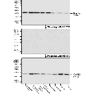
Figure 1. Representative blots for Neurofilament L of Hello Bio SuperBlotTM ECL Western Blotting Substrate Kit (Standard) and competitor solutions

Representative blots of Hello Bio ECL solutions and competitors showing SuperBlotTM ECL Western Blotting Substrate Kit (Standard) providing similarly clear blots to all competitors with low background. Method: A 12% acrylamide gel was loaded with a dilution series of rat brain cytosol fraction (10µg to 5ng total protein per lane) and transferred to a PVDF membrane. This was then probed with a monoclonal rabbit anti-neurofilament L antibody (HB7266, 1:10,000 dilution, 100ng/ml) primary antibody and goat anti-rabbit HRP (1:10,000 dilution) secondary antibody. The blot was exposed with every ECL solution (500µl part A + 500µl part B) using a Licor Odyssey Fc digital imager . For more information on Western Blotting please see our Western blotting protocol

Figure 2. Fully counterbalanced comparison of Hello Bio ECL substrates with competitor products.

SuperBlotTM ECL Western Blotting Substrate Kit (Standard) shows similar performance to Thermofisher Pierce ECL Western Blotting Substrate in Western Blotting. Method: 12% acrylamide gels were loaded with a dilution series of rat brain cytosol fraction (10µg to 5ng total protein per lane) and transferred to PVDF membranes. These were then probed with a monoclonal rabbit anti-neurofilament L antibody (HB7266, 1:10,000 dilution, 100ng/ml) primary antibody and goat anti-rabbit HRP (1:10,000 dilution) secondary antibody. All blots were exposed with every ECL solution (500µl part A + 500µl part B) in a fully randomised Latin square design to avoid any order effects. ECL exposure was carried out following the protocol detailed in "Application Notes". Imaging was carried out using a Licor Odyssey Fc digital imager and all band intensities were calculated in Licor Image Studio. For more information on Western Blotting please see our Western blotting protocol

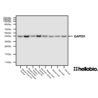
Figure 3. GAPDH Western Blot exposed using SuperBlotTM ECL Western Blotting Substrate Kit (Standard)

Western Blot with a range of tissue lysates and preparations probed for GAPDH with a mouse monoclonal anti-GAPDH antibody (HB9177) and developed using SuperBlotTM ECL Western Blotting Substrate Kit (Standard). Method: mouse brain and rat brain membrane (P2) and cytosol fractions were prepared following previous work (Molnar et al., 1993. Neuroscience 53:307-326) from freshly collected adult brains. Other tissue lysates were prepared following established protocols from freshly dissected tissue (see our guide on WB sample preparation). Samples were loaded (20µg / lane) onto a 4-20% acrylamide gel alongside a protein ladder (NEB Prestained) before being run at 150V for 50 minutes. Wet transfer to a PVDF membrane was completed in 90 minutes using 400mA. The membrane was blocked for 2hrs in 5% non-fat dry milk before being incubated overnight at 4°C in HB9177 at a 1:4,000 dilution. Following washing, the membrane was incubated in secondary antibody (1:10,000 dilution, Polyclonal goat anti-mouse HRP conjugated, Sigma, A3682) for 2hrs. For more detail please see our Western blotting protocol. Detection was accomplished using SuperBlotTM ECL Western Blotting Substrate Kit (Standard) and a Licor Odyssey Fc imaging system (ECL channel: 4.5 min exposure, 700nm channel: 30 sec exposure).

Figure 4. Histone H3 Western Blot exposed using Hello Bio SuperBlotTM ECL Western Blotting Substrate Kit (Standard).

Western Blot with a range of tissue lysates and preparations probed for Histone H3 with a rabbit polyclonal anti-histone h3 antibody (HB6980) and developed using SuperBlotTM ECL Western Blotting Substrate Kit (Standard). Method: mouse brain and rat brain membrane (P2) and cytosol fractions were prepared following previous work (Molnar et al., 1993. Neuroscience 53:307-326) from freshly collected adult brains. Other tissue lysates were prepared following established protocols from freshly dissected tissue (see our guide on WB sample preparation). Samples were loaded (20µg / lane) onto a 20% acrylamide gel alongside a protein ladder (BioRad Precision Plus Dual Colour) before being run at 60V for 30 minutes followed by 100V for 80 minutes then 130V for 40 minutes. Wet transfer to a PVDF membrane was completed in 90 minutes using 400mA. The membrane was blocked for 2hrs in 5% non-fat dry milk before being incubated overnight at 4°C in HB6980 at a 1:1,000 dilution. Following washing, the membrane was incubated in secondary antibody (1:10,000 dilution, Polyclonal goat anti-rabbit HRP conjugated, Sigma, A6154) for 2hrs. For more detail please see our Western blotting protocol. Detection was accomplished using Hello Bio ECL SuperBlotTM ECL Western Blotting Substrate Kit (Standard) and a Licor Odyssey Fc imaging system (ECL channel: 6 min exposure, 700nm channel: 30 sec exposure).

Biological Data

Application notes

Protocol for Chemiluminescent blot development with ECL

Digital Imaging

  1. Remove blot from the final wash solution and place on an imaging tray
  2. Mix equal quantities of part A and part B solutions being careful not to contaminate solutions by changing pipette tips
    1. For a 10x7.5cm gel we recommend 750µl of each solution
  3. Add combined solutions to the blot making sure to cover the entire area.
  4. Cover blot with a clear transparent sheet of plastic to prevent evaporation then immediately image.
    1. Be careful to not introduce any bubbles as these will show up in the final image.

Film Imaging

  1. Mix equal quantities of part A and part B solutions being careful not to contaminate solutions by changing pipette tips.
    1. For a 10x7.5cm gel we recommend 750µl of each solution
  2. Add combined solutions to a sheet of cling film large enough to fit the blot.
  3. Remove the blot from the final wash buffer, dab off any excess then place into the ECL solution. Use tweezers move the blot in order to soak it in ECL making sure to cover each side thoroughly.
  4. Dab off any excess ECL with filter paper then place into a pocket of clear plastic within a X-ray imaging cassette.
    1. Be careful to not introduce any bubbles as these will show up in the final image.
  5. Move to a dark room.
  6. Cut a piece of X-ray film to size then tape to the opposing door of the cassette. Close the cassette in one clean motion so that the film and blot are now in contact.
  7. Expose the film for an appropriate amount of time.
    1. This will vary depending on the target protein and which primary and secondary antibodies are used.
  8. Open the cassette and develop the film.

Solubility & Handling

Storage instructions

+4°C (protect from light)

Storage buffer

Contains 0.05% ProClin-300

Important This product is for RESEARCH USE ONLY and is not intended for therapeutic or diagnostic use. Not for human or veterinary use.

Calculators

Molarity

=
x
x
More Info

Dilution

x
=
x
More Info